Z-2159

ZytoLight® SPEC TP53/ATM Dual Color Probe
orange to green, pkg of 0.2 mL, pkg of 0.05 mL
Synonym(s):
FISH probe, dual color FISH probe
Sign In to View Organizational & Contract Pricing.
Select a Size
Change View
packaging
pkg of 0.05 mL, pkg of 0.2 mL, pkg of 20 tests, pkg of 5 tests
storage condition
protect from light
color
orange to green
application(s)
research use
storage temp.
2-8°C
General description
The ZytoLight® SPEC TP53/ATM Dual Color Probe is a direct-labeled FISH probe designed for the qualitative detection of deletions in the human TP53 and ATM genes in cytologic specimens using fluorescence in situ hybridization (FISH). It is optimized for use with the ZytoLight® FISH-Cytology Implementation Kit. ZytoLight® probes enable the precise identification of genetic abnormalities, including translocations, deletions, amplifications, and chromosomal aneuploidies, in formalin-fixed, paraffin-embedded (FFPE) tissue sections or cytology specimens, without the need for additional antibody-based detection.
Components
The ZytoLight SPEC TP53/ATM Dual Color Probe comprises:
Principle
The fluorescence in situ hybridization (FISH) method enables the identification and visualization of particular nucleic acid sequences within cell samples. DNA fragments labeled with fluorescent dyes, known as FISH probes, are co-denatured with their complementary target DNA strands in the sample and then allowed to hybridize. Following hybridization, non-specific and unbound probe fragments are removed through stringency washes. The DNA is then counterstained with DAPI, and the hybridized probes are detected using a fluorescence microscope equipped with filters matched to the specific fluorochromes used to label the FISH probes.
Components
The ZytoLight SPEC TP53/ATM Dual Color Probe comprises:
- ZyOrange (excitation 547 nm/emission 572 nm) labeled polynucleotides (~4.5 ng/µl), targeting sequences mapping in 17p13.1* (chr17:7,495,749-7,663,022) harboring the TP53 gene region;
- ZyGreen (excitation 503 nm/emission 528 nm) labeled polynucleotides (~10.0 ng/µl), targeting sequences mapping in 11q22.3* (chr11:107,957,618-108,380,921) harboring the ATM gene region;
- Formamide based hybridization buffer
Principle
The fluorescence in situ hybridization (FISH) method enables the identification and visualization of particular nucleic acid sequences within cell samples. DNA fragments labeled with fluorescent dyes, known as FISH probes, are co-denatured with their complementary target DNA strands in the sample and then allowed to hybridize. Following hybridization, non-specific and unbound probe fragments are removed through stringency washes. The DNA is then counterstained with DAPI, and the hybridized probes are detected using a fluorescence microscope equipped with filters matched to the specific fluorochromes used to label the FISH probes.
Application
This probe is intended for use in cytologic specimens to qualitatively detect human TP53 and ATM gene deletions, via FISH.
Analysis Note
Using suitable filter sets, the probe’s hybridization signals show as orange for the TP53 gene region and green for the ATM gene region.
- Normal scenario: in normal cells, or cells without deletions in these gene regions, two green and two orange signals are visible during interphase.
- Aberrant scenrio: if the TP53 gene region is deleted, fewer orange signals are present. Partial deletions of the TP53 locus may produce orange signals that are smaller but maintain the usual signal pattern. For deletions in the ATM gene region, fewer green signals are detected. Partial deletions in the ATM gene region may result in green signals of reduced size, while the overall signal pattern remains unchanged
Legal Information
ZytoLight is a registered trademark of ZytoVision GmbH in Germany
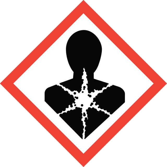
Signal Word
Danger
Hazard Codes
Precautionary Statements
Hazard Classifications
Carc. 2 - Repr. 1B - STOT RE 2 Oral
Target Organs
Blood
Storage Class
6.1C - Combustible acute toxic Cat.3 / toxic compounds or compounds which causing chronic effects
Flash Point (°F)
Not applicable
Flash Point (°C)
Not applicable
Choose from one of the most recent versions:
Already Own This Product?
Find documentation for the products that you have recently purchased in the Document Library.