
Z-2197

ZytoLight® SPEC PDGFRB Dual Color Break Apart Probe
Synonym(s):
FISH probe, dual color FISH probe
Select a Size
Change View
About This Item
eCl@ss:
32039090
product line
ZytoVision
packaging
pkg of 0.05 mL, pkg of 5 tests
manufacturer/tradename
ZytoVision
storage condition
protect from light
color
green to orange
application(s)
research use
storage temp.
2-8°C
General description
The ZytoLight® SPEC PDGFRB Dual Color Break Apart Probe (PL155) is a direct-labeled FISH probe for the qualitative detection of translocations of the human PDGFRB gene at 5q32 in cytologic specimens, using fluorescence in situ hybridization (FISH). The probe is intended for use with the ZytoLight FISH-Cytology Implementation Kit.
ZytoLight probes enable the precise identification of genetic abnormalities, including translocations, deletions, amplifications, and chromosomal aneuploidies, in formalin-fixed, paraffin-embedded (FFPE) tissue sections or cytology specimens, without the need for additional antibody-based detection.
Components
The ZytoLight SPEC PDGFRB Dual Color Break Apart Probe comprises:
Principle
The fluorescence in situ hybridization (FISH) method enables the identification and visualization of particular nucleic acid sequences within cell samples. DNA fragments labeled with fluorescent dyes, known as FISH probes, are co-denatured with their complementary target DNA strands in the sample and then allowed to hybridize. Following hybridization, non-specific and unbound probe fragments are removed through stringency washes. The DNA is then counterstained with DAPI, and the hybridized probes are detected using a fluorescence microscope equipped with filters matched to the specific fluorochromes used to label the FISH probes.
ZytoLight probes enable the precise identification of genetic abnormalities, including translocations, deletions, amplifications, and chromosomal aneuploidies, in formalin-fixed, paraffin-embedded (FFPE) tissue sections or cytology specimens, without the need for additional antibody-based detection.
Components
The ZytoLight SPEC PDGFRB Dual Color Break Apart Probe comprises:
- ZyGreen (excitation 503 nm/emission 528 nm) labeled polynucleotides (~10.0 ng/µl), targeting sequences mapping in 5q32-q33.1* (chr5:149,548,518-150,285,722) distal to the PDGFRB breakpoint region;
- ZyOrange (excitation 547 nm/emission 572 nm) labeled polynucleotides (~4.5 ng/µl), targeting sequences mapping in 5q32* (chr5:149,058,515-149,471,369) proximal to the PDGFRB breakpoint region;
- Formamide based hybridization buffer
Principle
The fluorescence in situ hybridization (FISH) method enables the identification and visualization of particular nucleic acid sequences within cell samples. DNA fragments labeled with fluorescent dyes, known as FISH probes, are co-denatured with their complementary target DNA strands in the sample and then allowed to hybridize. Following hybridization, non-specific and unbound probe fragments are removed through stringency washes. The DNA is then counterstained with DAPI, and the hybridized probes are detected using a fluorescence microscope equipped with filters matched to the specific fluorochromes used to label the FISH probes.
Application
For the qualitative detection of translocations involving the human PDGFRB gene at 5q32 by fluorescence in situ hybridization (FISH)
Analysis Note
Using suitable filter sets, the probe hybridization signals appear green away from the PDGFRB breakpoint region and orange near it.
- Normal scenario: in normal cells during interphase or cells without a PDGFRB gene region translocation, two green/orange fusion signals are observed.
- Aberrant scenario: one translocated PDGFRB gene region is shown by one separate green and orange signal each.
Legal Information
ZytoLight is a registered trademark of ZytoVision GmbH in Germany
Signal Word
Danger
Hazard Codes
Precautionary Statements
Hazard Classifications
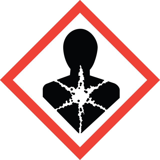
Carc. 2 - Repr. 1B - STOT RE 2 Oral
Target Organs
Blood
Storage Class
6.1C - Combustible acute toxic Cat.3 / toxic compounds or compounds which causing chronic effects
Choose from one of the most recent versions:
Already Own This Product?
Find documentation for the products that you have recently purchased in the Document Library.