Counterstaining after the Duolink In Situ Protocol
We recommend applying the counterstaining protocol after the completion of the Amplification step in section 7.3, step 5 of the Duolink In Situ Fluorescence User Manual. The following protocol exemplifies counterstaining using a directly labeled FITC anti-alpha tubulin antibody:
Perform all the steps at room temperature.
- Tap off the Amplification-Polymerase solution from the slides.
- Wash the slides in 1x Wash Buffer B for 2 x 10 min.
- Move the slides to 1x Wash Buffer A for 1 min
- Proceed to the counterstaining protocol and incubate the sample with e.g. a FITC labeled mouse anti-alpha tubulin for 40 min. - If the antibody used for counterstaining requires an antibody diluent different from that used during the Duolink assay, it is advised to add a blocking step before incubation with the counterstaining antibody
- Wash the slides with 1x Wash Buffer A for 2 x 2 min.
- Wash the slides with 0.01x Wash Buffer B for 1 min.
- Mount the slides following the protocol in section 7.3, step 7 in the Duolink In Situ Fluorescence User Manual.

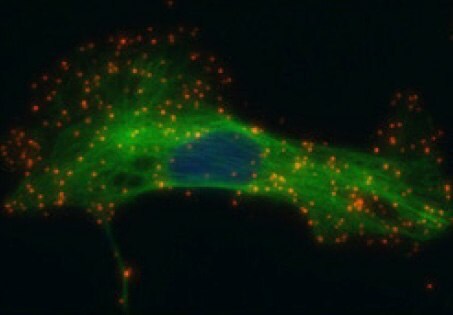
Above:Single recognition of HER2 in SK-BR-3 cells. Red: PLA signals, each representing one HER2 protein. Green: counterstain of alpha tubulin. Blue: nucleus. PLA probes anti-Rabbit PLUS and MINUS, Duolink In Situ Detection Reagents Orange.
Custom Services
Does your experiment require additional customization? Let us do the work for you. Learn more about our Custom Services program to accelerate your Duolink® PLA projects.
If you would like to receive additional information on Duolink, contact us at (800) 325-5832.
To continue reading please sign in or create an account.
Don't Have An Account?