Residual Peroxide Determination in Food Packaging
The extracellular (ECM) microenvironment, defined by biochemical cues and physical cues, is a deciding factor in a wide range of cellular processes including cell adhesion, proliferation, differentiation, and expression of phenotype specific functions. For this reason, engineering the ECM microenvironment provides clear benefits in studies of cell and tissue engineering and related applications. Currently existing technology offers simple and merely adequate environments that facilitate simple cell processes such as cellular adhesion.1,2 The simple presentation of cell adhesion motifs is not optimal for controlling more integrated processes.
Crosstalk among signaling pathways act synergistically to enhance cellular responses such as cell adhesion or proliferation3. A recent study showed that a combination of extracellular matrix derived peptides presented on a surface may enhance cell adhesion strength and focal adhesion assembly. The combinatorial presentation of ECM peptides on cell growth surfaces may also promote elevated proliferation rates of primary or stem cells4,5,6.
The Kollodis ECM Library provides a means to regulate a variety of cell surface receptors for your cell studies with the highlighted features provided below.
Advantageous Features
- Offers a wide range of ECM derived peptide containing mussel adhesive proteins based casting to meet the needs of many cell culture systems
- Flexible engineering of biochemically defined surfaces to specifically direct your cell by manipulating your cell surface receptors
- Easy-to-use, in situ formable 3D MAPTrix HyGel™ matrix microenvironment
- Ensures consistently high-quality products with extensive quality control (QC) testing
Collagen derived peptide
Collagens serve as scaffolds for the attachment of cells and matrix proteins; but, they are increasingly being recognized to have many other ligands and to be highly biologically active. For example, collagens provide integrin binding motifs and heparin binding motifs. The α2β1 integrin recognizes GXO/SGER such as GFPGER or GFOGER, and these sites are for endothelial cell binding and activation, and for angiogenesis. Integrin binding sites for αvβ3 have antitumor activity, and may inhibit the activation of human neutrophil or the proliferation of capillary endothelial cells. Integrin binding sites in the NC1 domains have anti-angiogeneic properties mediated by the α1β1 or αvβ3 integrin binding.

Figure 1.Collagen Derived Peptide
Fibronectin derived peptide
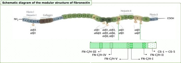
An ECM Mimetic Library for Engineering Surfaces to Direct Cell Surface Receptor Binding Specificity and Signaling Laminin derived peptide (α1 ~ α5, β, γ chains) Fibronectin naturally exists as a dimer, consisting of two nearly identical monomers. Two regions in each fibronectin subunit possess cell binding activity: III9-10 and III14-V (refer to the modular structure of fibronectin below). The primary receptor for adhesion to fibronectin commonly involves the RGD motif of repeat III10 through integrins such as α5β1; however, this integrin-ligand interaction is only sufficient for cell attachment and spreading. Additional signaling through the cell surface proteoglycan such as syndecan-4 is required for focal adhesion formation and rearrangement of the actin cytoskeleton into bundled stress fibers. This binding occurs primarily via the HepII domain (containing the FN type III repeats 12-14) in the C-terminal region of fibronectin.

Figure 2.Fibronectin Derived Peptide
Laminin Derived Peptide (α1 ~ α5, β, γ Chains)
Laminins, heterotrimers composed of α, β, and γ chains, are multifunctional glycoproteins present in basement membranes. Integrins, dystroglycan, syndecans, and several other cell surface molecules are cellular receptors for laminins. The globular domains located in the N- and C-terminus of the laminin alpha chains are critical for interactions with the cellular receptors. Integrin α6β1 binds to most of the laminin isoforms. Integrin α3β1 interacts with laminin-5 and -10/11 more specifically than the other isoforms. Integrins α1β1, α2β1, and α7β1 show binding activity to laminin-1 and -2. Interaction of integrin α6β4 with laminin-5 forms hemidesmosomes in the skin. α-dystroglycan strongly binds to the laminin α1 and α2 chains and moderately interacts with the α5 chain.

Figure 3.Lamanin Derived Peptide
Additional ECM-derived peptides
Cadherins are calcium dependent cell adhesion proteins which are involved in many morphoregulatory processes including the establishment of tissue boundaries, tissue rearrangement, cell differentiation, and metastasis. The extracellular domain of E-cadherin tends to bind in a homophilic manner; however, heterophilic binding occurs under certain conditions. The binding of extracellular cadherin is the basis for cell-cell adhesion and tends to be prevalent at adherins junctions and is structurally associated with actin bundles.
Other sets of extracellular matrix components - for example, vitronectin, nidogen or Tenascin, and SIBLINGs (small integrin-binding ligand, Nlinked glycoprotein) such as bone sialoprotein (BSP) or osteonpontin derived ligand - can also influence the cellular behavior by regulating cell signaling (directly or indirectly). Unlike the main extracellular matrix components such as collagen or fibronectin, these other proteins are adhesionmodulatory extracellular matrix proteins which interact with the main ECM components or integrins.
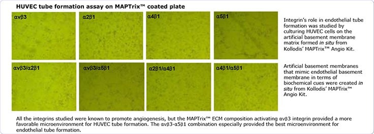
Kollodis BioSciences’ MAPTrix™ Technology provides a true ECM microenvironment by presenting combinatorial peptide motifs to induce and/or regulate combinatorial integrin mediated signaling for integrated cellular processes as demonstrated in endothelial tube formation on MAPTrix HyGel™ coated plates, as provided below.

Figure 4.ECM Derived Peptides
References
To continue reading please sign in or create an account.
Don't Have An Account?