manufacturer/tradename
Novagen®
storage condition
OK to freeze
shipped in
wet ice
General description
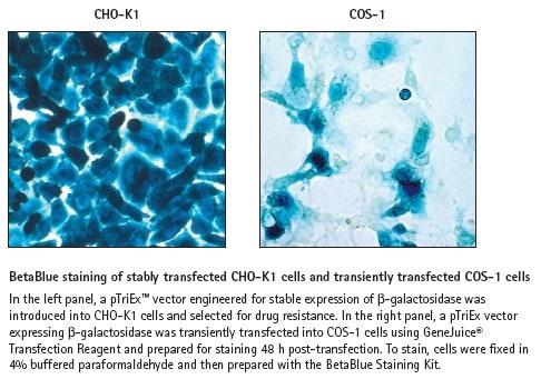
Convenient visualization of β-gal in cells or tissues
The BetaBlue
Other Notes
Legal Information
NOVAGEN is a registered trademark of Merck KGaA, Darmstadt, Germany
Disclaimer
Toxicity: Multiple Toxicity Values, refer to MSDS (O)
存储类别
10 - Combustible liquids