
biological source
mouse
Quality Segment
antibody form
purified antibody
antibody product type
primary antibodies
clone
34E4, monoclonal
form
lyophilized
contains
≤0.1% sodium azide as preservative (antibody only)
species reactivity
human
manufacturer/tradename
Calbiochem®
storage condition
OK to freeze, avoid repeated freeze/thaw cycles
dilution
(ELISA (0.1 µg/mL)
Immunoblotting (0.5 µg/mL, chemiluminescence))
isotype
IgG1
shipped in
wet ice
storage temp.
−20°C
target post-translational modification
phosphorylation (pSer374)
General description
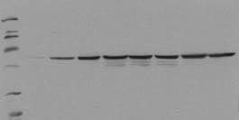
Mouse monoclonal antibody purified from serum-free cell culture supernatant by thiophilic adsorption and size exclusion chromatography. Supplied with a positive control lysate consisting of EGF-treated HepG2 cells. Recognizes the ~55 kDa c-fos protein phosphorylated at Ser374 by MAP kinase.
Recognizes the ~55 kDa c-Fos protein phosphorylated at Ser374. Does not detect the unphosphorylated form.
The early gene product c-Fos is expressed following mitogenic stimulation and functions as a sensor for MAPK signal duration. When MAPK activation is transient, MAPK activity declines before accumulation of the c-Fos protein. When MAPK activation is sustained, c-Fos is phosphorylated by MAPK at Ser374. Phosphorylation stabilizes the Fos protein and primes c-Fos for additional phosphorylation at Thr325.
This PhosphoDetect Anti-Fos (pSer³⁷⁴) Mouse mAb (34E4) is validated for use in ELISA, Immunoblotting for the detection of Fos (pSer³⁷⁴).
Immunogen
Human
a synthetic phosphopeptide corresponding to amino acids surrounding the Ser³⁷⁴ phosphorylation site of human c-Fos
Application
ELISA (0.1 g/ml)
Immunoblotting (0.5 g/ml, chemiluminescence)
Physical form
100 µg antibody lyophilized from 2X PBS, PEG, sucrose and 200 µl lyophilized control lysate from EGF-treated HEPG2 cells.
Preparation Note
Reconstitute antibody with 1 ml water (15 minutes, room temperature). Following reconstitution, aliquot and freeze in liquid nitrogen. Reconstituted antibody can be stored at -80°C for up to 1 year. Aliquots may be stored at 4°C for up to 3 months and should be thawed at 37°C. Reconstitute control lysate with 200 µl H₂O. After complete solubilization of the proteins add 200 µl SDS-PAGE sample buffer and incubate at 90°C for 5 min. Following reconsitution aliquot and freeze (-20°C). Avoid freeze/thaw cycles.
Analysis Note
Positive Control
HepG2 cells treated with EGF
HepG2 cells treated with EGF
Other Notes
Chen, R-H., et al. 1993. Proc. Natl. Acad. Sci. USA90, 10952.
Does not detect the unphosphorylated form of c-fos. For immunoblotting use 20 µl control lysate per lane (minigel) for chemiluminescent detection. Antibody should be titrated for optimal results in individual systems.
Legal Information
CALBIOCHEM is a registered trademark of Merck KGaA, Darmstadt, Germany
Disclaimer
Toxicity: Irritant (B)
Still not finding the right product?
试用我们的 产品选型工具 工具缩小选择范围
存储类别
11 - Combustible Solids