Microscopic Examination of Samples
Methodes of Microscopic Examination of Samples
1. Viewing from below the plate (through transparent PET or CM membranes)
Millicell® devices using PET or CM membrane have been designed to allow visualization of cells from below using an inverted microscope. For viewing live cells, microscopic observations can be made through the receiver or plastic plate containing media. In order to focus on the cells, the microscope objective (typically 5–20X) must have an appropriate working distance. (For objective specifications, visit the websites listed in the Microscope Objective Information section.) Fixed cells that do not require to be visualized in media can be viewed directly without the receiver plate. However, care should be taken not to contaminate the objective with liquid residue (media, mounting fluid) on the membrane.
2. Viewing from above the plate (Millicell® inserts or Millicell®-24 cell culture insert plates)
Some cell culture platforms can allow the cells to be viewed in a conventional microscope directly from above using low magnification. Cells can be visualized through the lid to maintain sterility or with the lid removed for fixed cells or when maintenance of sterility is not required. Working distances of the objective must be longer when reading from above compared to when reading from below. If using immunofluorescence, it is recommended to use a mounting fluid that contains an anti-fade additive to prevent photobleaching.
3. Visualizing membranes on microscope slides (for higher magnification or with objectives with short working distances [less than 2 mm])
The membrane can be removed from each well for microscopic evaluation. This allows for higher magnification examination and storage of the slides for future use.

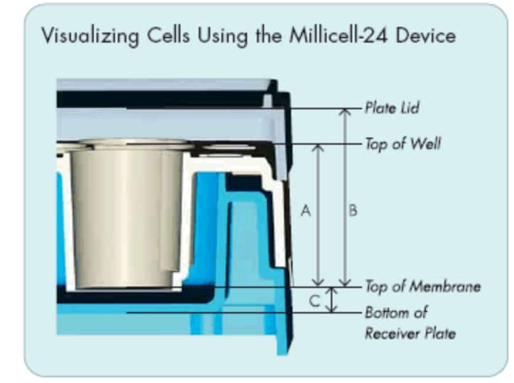
Figure 1. Visualizing Cells using the Millicell-24 Device
For visualizing from above the membrane, typically 5–20X objectives are used that have at least a 13.59 mm (A) or a 18.03 mm (B) working distance when viewing without or with the lid, respectively. For visualizing from below the membrane, 5–20X objectives are used that have at least a 2 mm (C) working distance.
Preparation of Membranes on Microscope Slide
- Remove the membrane from the well using a sharp scalpel to make a small incision in the edge of the membrane. Carefully cut along the inside of the well wall for approximately one quarter of the well diameter. Using forceps (Product No. XX6200006P), carefully hold the membrane while continuing to cut around the well diameter to remove membrane. Alternatively, a cork borer may be used to remove the membrane. Note: Use care to prevent membrane from curling.
- Place the membrane disk, cells facing up, onto a microscope slide.
- Add 50 µL mounting fluid to the membrane disk and allow it to wet out in order to prevent bubbles under the disk.
- Slowly lower a cover slip onto the membrane at an angle to allow air bubbles to be removed.
Microscope Objective Information
Information regarding microscope objective magnification power and working distances can be obtained from individual optical dealers or from the microscope vendors:
- Nikon Instruments
- Olympus Corporation
- Carl Zeiss
NoteIt is assumed that users of this procedure will be knowledgeable in TEM procedures.
Materials and Reagents
- Millicell®Cell Culture Inserts
- Phosphate Buffered Solution (PBS)
- Glutaraldehyde
- Osmium tetroxide
- Sodium cacodylate
- Sucrose, reagent grade
- Calcium chloride
- Lead nitrate
- Sodium citrate/Sodium hydroxide
- Uranyl acetate
- Ethanol
- Flat embedding trays
- Embedding Resin (i.e. EPON812)
- Fine forceps (Product No. XX6600006P)
- Diamond Knife
- Cork Borer
- Durapore Filter Disk — Millipore
Sample Pictures

Figure 2. Living murine embryonic stem cell derived embryoid bodies visualized in a 1 um PET Millicell®-24 device using an Olympus IMT-2 inverted microscope

Figure 3. Neuron differentiation of embryonic stem cells in Millicell®-24 1 um PET filter plates.
Murine embryonic stem cells were formed into suspended embryoid bodies (EBs), then transferred to 1 um PET Millicell®-24 plates for attachment and differentiation. The photo inset shows the inverted phase contrast through membrane of live EBs in the media. Neural differentiation after netinoic acid treatment of attached EBs was confirmed by anti-neurofilament immunofluoresence.
A. Processing/Cell Preparation
Note: Steps 1–5 should be done on an intact Millicell®cell culture insert or plate well.
- Wash cells briefly (2 times for 5 minutes each) at room temperature with phosphate buffered solution without fixative.
- Fix cells in 2% glutaraldehyde in 100 mm sodium cacodylate buffer, pH 7.5, at room temperature from 15 minutes to 2 hours.
- Wash cells (2 times for 5 minutes each) in 100 mm sodium cacodylate buffer at room temperature. Note: At this point, cells can be stored in the above buffer with 7 g sucrose/100 mL buffer at 4 °C.
- Fix cells in 1% osmium tetroxide in either 100 mm sodium cacodylate or suitable phosphate buffer.
- Dehydrate cells in the following concentrations of ethanol:
- For infiltration, EPON812, an EDPON substitution, or LX112 is suitable for both devices (do not use Spurr’s). The following is a general infiltration scheme:
Note: It is not necessary to use any other agent, such as propylene oxide, with plastic. Propylene oxide will dissolve the cellulosic filters. In addition, the standard inversion/rotation of specimens used in these steps is not advised since either (1) damage to the cell layer or (2) stretching of the cellulosic filter may occur. Mild shaking on a gel shaker apparatus is sufficient for successful infiltration.
Note: Before the next step the membrane must be detached from the surrounding plastic ring. Sometimes this will occur without manipulation since the EPON may loosen the membrane-to-ring bond. If this does not occur, use a sharp scalpel or a cork borer and cut the membrane. It may also help to cut the membrane over a 47 mm filter support disk. Under no circumstances should the membrane be left attached to the ring during polymerization.
- Transfer to fresh plastic and polymerize at 68 °C overnight.
B. Sectioning Notes
- Nitrocellulose (HA), polycarbonate (PC) and polyethylene terepthalate (PET) membrane: These membranes can be sectioned in any plane without difficulty.
- CM (Biopore) membrane: The Biopore membrane must be processed in one of two ways based on the final thickness of the section.
Materials
To continue reading please sign in or create an account.
Don't Have An Account?