High purity water for DNA microarrays
DNA microarray analysis involves multiple steps, including oligonucleotide chemistry, hybridization, washing, and fluorescence detection, that are extremely sensitive to trace contaminants. Poor water quality can compromise data quality, reproducibility, and sensitivity of the technology.
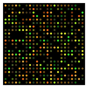
Overview of DNA microarrays
DNA microarrays, also referred to as gene chips or DNA chips, contain many tiny spots bound to a solid support (e.g., glass, silicon chip), with each spot holding multiple copies of a specific gene sequence. Spot sizes vary depending on the way the microarray is manufactured, but they are usually less than 200 µm in diameter with each spot containing picomoles of a specific DNA sequence. A single microarray can contain up to tens of thousands of spots consisting of DNA, cDNA or oligonucleotides.
Microarrays can be manufactured in different ways, depending on the number of spots/targets, costs, customization requirements, and the type of scientific question being asked. Arrays may have as few as 10 targets to up to 2.1 million µm-scale targets.
Principle of DNA microarrays
To analyze a sample using DNA microarray, the nucleic acids must first be extracted and purified, converted to cDNA or cRNA, and labeled with a detectable marker, such as a fluorescent dye, silver, or chemiluminescent tag. The labeled nucleic acids are then applied to the microarray, and any nucleic acids that are complementary to the microarray probes will bind, while non-binding nucleic acids are removed by washing. Bound nucleic acids can be quantified based on relative abundance.
Multiple samples can be applied to a DNA microarray at once using a different tag for each sample. For example, cDNA from a normal cell and cDNA from a pathological cell can be labeled with different fluorescent tags. The resulting signal detected can distinguish between the presence of a specific sequence in both cells (yellow dots), only one cell type (red or green dots), or neither cell (grey dots) (Figure 1).
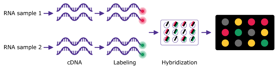
Figure 1.DNA microarray experimental workflow.
Application of DNA microarray
DNA microarrays have revolutionized the field of genomic research by providing a robust, high-throughput platform for understanding complex genetic interactions and their effects on health, development and disease. Arrays have many applications across multiple fields such as genetics, oncology, pharmacogenomics and environmental science.1-4
Gene expression profiling
Microarrays are extensively used to measure the expression levels of numerous genes at once to understand gene activity across different conditions, treatments, time periods, or developmental states. This application is crucial in identifying which genes are turned on or off, for example, in different stages of a disease, in response to a drug, or under stress conditions in plants and animals. Microarray technology can be used to develop specific gene expression profiles for particular diseases, especially cancers. Comparing the gene expression profiles of healthy vs. diseased tissues can help in diagnosing the type of cancer and its progression.
Methylation profiling
Methylation profiling, or DNA methylation profiling, measures methylation patterns across CpG sites. CpG sites are positions in the genome where a cytosine is followed by a guanine, and the cytosine may be methylated. It works using a conversion step (e.g. bisulfite treatment) to distinguish between methylated and unmethylated cytosines, followed by microarray analysis to identify methylated sites.5 Methylation profiling reveals signatures that can be used to diagnose or classify diseases like cancer.
Comparative genomic hybridization (CGH)
Cancer and genetics research use microarrays to detect and map gene copy number variations across different samples. This is particularly useful in oncology to identify genomic gains and losses associated with various cancers.
Genotyping and genetic mapping
Microarrays can be used to detect Single Nucleotide Polymorphisms (SNPs) across the genome, aiding in genotyping, mapping genetic diseases, and identifying genetic predispositions to various conditions such as response to drugs, susceptibility to diseases, and physical traits.
Pathogen detection
Microarrays can be designed to detect a wide range of clinical and environmental pathogens (bacterial, viral, fungal) by checking for the presence of pathogen-specific DNA sequences. This application is essential in quickly diagnosing infections, monitoring outbreaks, and in biosecurity.
Pharmacogenomics
Personalized medicine has advanced significantly due to genomic microarrays. By analyzing how genes affect an individual's response to drugs, microarrays can help tailor medical treatment to patients' genetic makeup, maximizing therapeutic effects and minimizing adverse effects.
Environmental sciences
Microarrays are used in ecotoxicology to assess the health of an ecosystem. By analyzing gene expression changes in organisms exposed to environmental stressors such as pollutants, arrays provide insight into the biological impact of environmental contaminants.
Agricultural applications
Microarrays are used in crop improvement to study stress responses like drought, heat and salinity. Microarrays can be used as large screens to identify stress-resistant genes, which can help in breeding more resilient crop varieties.
Use of water in DNA microarrays
- Water is used during the microarray manufacturing process. Whether the technique selected is photolithography, printing or electrochemistry, rinsing steps are always necessary, requiring a significant amount of high purity water. Photolithography and electrochemistry techniques also require performing chemical reactions to build and grow the oligonucleotide sequences on the solid surface. This step also requires buffers that must be prepared with high purity water.
- Microarray experiments use water at nearly every step. First, nucleic acid extraction and PCR-based DNA amplification techniques [e.g. simple PCR or reverse transcription PCR (RT-PCR)] used to prepare the DNA require nuclease-free high purity water (see article, Water for PCR Techniques). Then, hybridization of the target nucleic acid to the probe requires a water-based buffer. A washing step that removes unspecific binding on the solid surface also requires buffers.
Water quality required for DNA microarrays
The high-purity water selected should comply with the various steps and materials used throughout the DNA microarray experiments. Water used to manufacture microarrays and, in the experiment, share some common quality requirements.
Nucleases
Water used for DNA microarrays should be nuclease free to prevent nuclease degradation of the sample, purified nucleic acids, and the microarray probes. Nuclease-free water is recommended during the nucleic acid extraction and the DNA amplification steps.6 Nuclease removal is efficiently done using ultrafiltration. A point-of-use ultrafiltration cartridge can be installed at the outlet of water purification systems to provide nuclease-free water on demand. For more information on preparing nuclease-free water, see the article Water for Nuclease-sensitive Molecular Biology Studies.
Organics
Organics could generate issues during the preparation of microarrays. In the printing processes, the solid surface must be perfectly clean to optimize printing. The presence of organic contaminants at concentrations as low as 5 ppb reduces the efficiency of oligonucleotide adhesion onto the surface. In other fabrication processes, where the DNA reacts with the surface of the slide, organics from the water could react non-specifically with the chemical sites of the surface, reducing the chance of reaction with the oligonucleotide. Organic molecules could also create interferences in the hybridization process.
Organic contaminants in water have an impact on the detection step (be it by fluorescence or chemiluminescence). Some organics can generate or quench fluorescence, and interfere with fluorescent signal detection, so a low level of organics also optimizes the detection step.
Ions
The concentration of ions is important during the hybridization step. Starting with water virtually free of ions (resistivity 18.2 MΩ•cm) ensures the preparation of buffers at the right ionic strength and pH.
In addition, polymerases used for PCR need a specific magnesium concentration for maximum efficiency and are susceptible to inhibition with metals such as cadmium and iron. Therefore, metal-free water should be selected for PCR-based DNA amplification. For more information on the impact of water contaminants on PCR, refer to the relevant section in the article, Water for PCR Techniques.
Silica
Silica can create a film on the solid surface, reducing the efficiency of the printing process. Water with low silica levels is, therefore, highly recommended. Different purification technologies, such as reverse osmosis, electrodeionization and ion exchange resins, are usually combined to remove silica from water.
Ozone
Cy5 fluorescence is highly sensitive to oxidative degradation. Reactive oxygen species (ROS), especially ozone, are well-established contributors to the fading of Cy5 signals. Ozone does not come from the water purification process, but it is present in the air and dissolves in water when water is left standing openly in air. Issues due to ozone may be reduced by handling high purity water away from sources of ozone, such as ozone-generating sterilizers, and reducing storage time of purified water.
Impact of water quality on DNA chips and microarrays
We compared DNA microarrays prepared with different grades of ultrapure water (Figure 2). Both preparations used dual-labeling with Cy3 and Cy5 fluorescent dyes.
- Experiment A: Water was prepared using a combination of reverse osmosis (RO) and ion exchange resins.
- Experiment B: Water was prepared with a system similar to the Milli-Q® IQ 7005 system, containing a combination of RO, electrodeionization (Elix® technology), activated carbon, UV photooxidation and ion exchange resins.
Both waters had resistivity of 18.2 MΩ•cm, ensuring low levels of ions, and bacteria levels below 1 cfu/mL. The level of organics (total organic carbon, TOC) differed between the two:
- Experiment A: TOC was 22 ppb (µg/L). This organic contamination could have negatively affected one or more steps in the experiment, causing a haze and poorer DNA microarray readings.
- Experiment B: TOC was 3 ppb. This lower TOC level yielded well-defined spots, a lower background and overall good results.
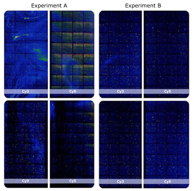
Figure 2.Comparison of dual-labelled DNA chips obtained using two different water qualities.
In summary, ultrapure water (resistivity 18.2 MΩ•cm) with low TOC (< 5 ppb) and low bacteria (< 0.1 cfu/mL) is suitable and recommended for DNA microarrays.
Ultrapure water for reliable and consistent DNA microarrays
Ultrapure (Type 1) water with low TOC level is essential for preparing and running DNA microarrays with minimal contamination, improved signal-to-noise ratio, and enhanced assay reproducibility. A range of water purification solutions adapted to the needs of scientists working with genomic microarrays is available.