Bacteriology

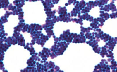
The discipline of bacteriology is concerned with all aspects of genetics, structure, physiology, behavior, pathogenicity, ecology, and evolution of bacterial species. Bacteriological investigations are essential in clinical diagnostics and industrial quality control. Application of microscopy in bacteriology involves the staining of microorganisms with suitable methods (e.g. Gram stain) to determine the Bacteria classification or to detect mycobacteria.
Featured Categories
We offer a complete selection of microscopy reagents, tools, and glassware for research and clinical lab analysis from Corning®, BRAND®, PELCO®, and other reliable specialty brands.
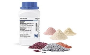
Discover high-quality microbial culture media. Choose from dehydrated or ready-to-use options, meeting industry standards and regulatory requirements.
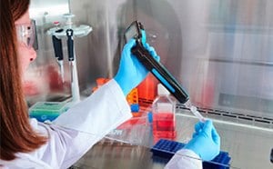
Accurate, consistent cell counting is critical for cell culture and downstream cellular assays and experiments. A handheld coulter counter provides the most accurate and reproducible cell counts in seconds. Disposable hemocytometers reduce risk in infectious settings.
Accurate, consistent cell counting is critical for cell culture and downstream cellular assays and experiments. A handheld coulter counter provides the most accurate and reproducible cell counts in seconds. Disposable hemocytometers reduce risk in infectious settings.
Gram Staining
Gram staining is an essential staining technique in bacteriology used to differentiate Gram-positive and Gram-negative bacteria. In this method, differentiation is achieved as Gram-positive bacteria retain a crystal violet stain due to the presence of a thick layer of peptidoglycan in their cell walls.
Alternatively, Gram-negative bacteria are decolorized by organic solvent and turn orange-pink when counterstained, which is attributed to a thinner peptidoglycan wall. Reagents required for this multi-step staining process include crystal violet (primary stain), aniline dye, iodine solution (mordant), and safranin orange (secondary stain), or carbol fuchsin counterstains.
Mycobacteria Staining
Early diagnosis of mycobacterial infection is critical, as the acid-fast bacteria are highly pathogenic and responsible for serious diseases, like tuberculosis. Diverse staining solutions are available for the detection of these pathogenic bacteria in histological tissue cultures and bacteriological smears. Fluorescence detection methods are utilized with the Ziehl-Neelsen staining technique, via either heat-treated slides (hot staining) or non-heated treatment (cold staining). This differential staining technique uses the lipid-soluble phenolic compound carbol fuchsin as the primary stain, and the counterstain, malachite green.
Trichomonad Staining
Trichomonas vaginalis parasites are especially common in gynaecological material, such as vaginal smears and urine sediment, and cause the most common non-viral sexually transmitted disease, trichomoniasis. Various stains like Giemsa and Acridine Orange, are used along with wet mount examination for the microscopic diagnosis of Trichomonas vaginalis.
Bacteriology applications in other industries
In the food and beverage industry, lactic acid bacteria such as Lactobacillus, Lactococcus, and Streptococcus are used in the manufacture of dairy products, such as cheese, buttermilk, and yogurt. Bacterial fermentations are used in the processing of beverages, such as tea and coffee. In the growing field of gut microbiome health, several bacterial species are used in probiotic supplements to reduce inflammation and improve bowel function. Bacteria are also used in the pharmaceutical industry in vaccine research and production, including tetracyclines, erythromycin by Streptomyces and bacitracin by Bacillus.
Visit our document search for data sheets, certificates and technical documentation.
Related Articles
- Information about lactobacilli, rod-shaped, Gram-positive, fermentative, facultative anaerobic or microaerophilic organotrophs. The lactobacillus organtroph belongs to the lactic acid bacteria group.
- Chromogenic media enable the selective detection of S. aureus, which produce bluish-green colonies that are clearly differentiated from other species.
- Selective media enable faster results and visual confirmation for the detection, identification, and enumeration of microorganisms
- Ribosomally synthesized antimicrobial peptides are a promising focus in antibiotic research amidst bacterial resistance and emerging infectious diseases.
- Common Cell Culture Problems: Contamination is easily the most common problem encountered in cell culture laboratories, sometimes with very serious consequence.
- See All (16)
Related Protocols
- Cell culture protocol for testing cell lines for bacterial and fungal contamination. Free ECACC handbook download.
- Initiating a Starter Culture
- General protocols for growth of competent cells in microbial medium.
- Proper laboratory glassware cleaning techniques are crucial for high-quality results in experiments and research.
- Lipopolysaccharides (LPS) are characteristic components of the cell wall of Gram negative bacteria; they are not found in Gram positive bacteria.
- See All (9)
Find More Articles and Protocols
How Can We Help
In case of any questions, please submit a customer support request
or talk to our customer service team:
Email custserv@sial.com
or call +1 (800) 244-1173
Additional Support
- Chromatogram Search
Use the Chromatogram Search to identify unknown compounds in your sample.
- Calculators & Apps
Web Toolbox - science research tools and resources for analytical chemistry, life science, chemical synthesis and materials science.
- Customer Support Request
Customer support including help with orders, products, accounts, and website technical issues.
- FAQ
Explore our Frequently Asked Questions for answers to commonly asked questions about our products and services.
To continue reading please sign in or create an account.
Don't Have An Account?


