technique(s)
cell culture | stem cell: suitable
flow cytometry: suitable
Quality Level
fluorescence
λex 488 nm
λex 532 nm
λem 615 nm
detection method
fluorometric
shipped in
wet ice
Related Categories
General description
Learn more about the AldeRed in Nature Communications! (Learn Now!)
High ALDH activity serves as a universal marker of stem cells, both normal and malignant. Cells can be identified and isolated based upon the enzymatic activity of ALDH, a detoxifying enzyme responsible for oxidation of hazardous aldehyde byproducts. The marker ALDH has been used to isolate cancer stem cells from various human malignancies including bladder, breast, cervical, colon, head and neck, liver, lung, pancreas, prostate and ovary. The AldeRed assay employs a fluorescent and non-toxic ALDH substrate (AldeRed 588-A) that diffuses freely into intact and viable cells, but remains trapped inside the cells once converted by ALDH into the corresponding acid. The amount of fluorescence produced is proportional to the ALDH activity in the cells and is measured by flow cytometry, allowing fluorescence-activated cell sorting (FACS). This kit supplies the ALDH inhibitor diethylaminobenzaldehyde (DEAB), which is used in negative control testing necessary for background fluorescence assessement.
Application
• All live intact cells (even with low ALDH expression) become fluorescent upon exposure to activated AldeRed 588-A. The generation of a DEAB-treated control is necessary for each cell sample to establish baseline fluorescence.
• Handle all reagents using aseptic techniques.
Stem Cell Research
Apoptosis & Cancer
Components
2. DEAB Reagent: (Part No. CS216595). One (1) vial containing 1 mL of DEAB (Diethylaminobenzaldehyde) reagent.
3. Hydrochloric Acid (2N): (Part No. CS216594). One (1) vial containing 1.5 mL of 2 Normal hydrochloric acid.
4. Dimethylsulphoxide: (Part No. CS216593). One (1) vial containing 1.5 mL of Dimethylsulphoxide (DMSO).
5. AldeRed Assay Buffer: (Part No. CS216592). Four (4) bottles containing 25 mL of AldeRed Assay Buffer each.
6. Verapamil: (Part No. CS220012). Four (4) vials containing 615 µg Verapamil powder each.
Quality
• AldeRed® Reagent is 95% pure (PMR)
Storage and Stability
• Reconstituted and activated AldeRed Reagent may be stored in aliquots at -20°C protected from light for up to 1 year. Repeated freezing and thawing is not recommended.
• AldeRed Assay Buffer supplemented with Verapamil may be stored at 2-8°C in aliquots for up to 3 months.
Legal Information
Disclaimer
Signal Word
Danger
Hazard Statements
Precautionary Statements
Hazard Classifications
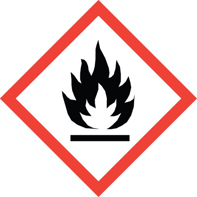
Acute Tox. 3 Oral - Aquatic Chronic 2 - Eye Irrit. 2 - Flam. Liq. 2 - Met. Corr. 1
Storage Class Code
3 - Flammable liquids
WGK
WGK 3
Flash Point(F)
55.4 °F
Flash Point(C)
13 °C
Regulatory Information
Certificates of Analysis (COA)
Search for Certificates of Analysis (COA) by entering the products Lot/Batch Number. Lot and Batch Numbers can be found on a product’s label following the words ‘Lot’ or ‘Batch’.
Already Own This Product?
Documents related to the products that you have purchased in the past have been gathered in the Document Library for your convenience.
Difficulty Finding Your Product Or Lot/Batch Number?
How to Find the Product Number
Product numbers are combined with Pack Sizes/Quantity when displayed on the website (example: T1503-25G). Please make sure you enter ONLY the product number in the Product Number field (example: T1503).
Example:
Additional examples:
705578-5MG-PW
PL860-CGA/SHF-1EA
MMYOMAG-74K-13
1000309185
enter as 1.000309185)
Having trouble? Feel free to contact Technical Service for assistance.
How to Find a Lot/Batch Number for COA
Lot and Batch Numbers can be found on a product's label following the words 'Lot' or 'Batch'.
Aldrich Products
For a lot number such as TO09019TO, enter it as 09019TO (without the first two letters 'TO').
For a lot number with a filling-code such as 05427ES-021, enter it as 05427ES (without the filling-code '-021').
For a lot number with a filling-code such as STBB0728K9, enter it as STBB0728 without the filling-code 'K9'.
Not Finding What You Are Looking For?
In some cases, a COA may not be available online. If your search was unable to find the COA you can request one.
Articles
AldeRed™ 588-A is a red fluorescent live cell probe that detects ALDH activity used to identify cancer stem cells and progenitor cells in culture. Cancer stem cells (CSCs) are subpopulations of cancer cells that can self-renew, generate diverse cells in the tumor mass, and sustain tumorigenesis. Some researchers believe that cancer arises from cancer stem cells that originate as a result of mutational hits on normal stem cells.
Read types of stem cells including multipotent stem cells, Pluripotent stem cells and iPSCs and their applications in basic stem cell research, stem cell therapy and disease modelling
Cancer stem cell media, spheroid plates and cancer stem cell markers to culture and characterize CSC populations.
Protocols
Step-by-step tumorpshere formation protocol that can used to generate 3D cancer stem cell spheroids.
Our team of scientists has experience in all areas of research including Life Science, Material Science, Chemical Synthesis, Chromatography, Analytical and many others.
Contact Technical Service


