Merck
CN
HomeStem Cell CultureCancer Stem Cells: Targets for Cancer Therapy

Cancer Stem Cells: Targets for Cancer Therapy

The Cancer Stem Cell Hypothesis

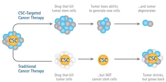
Are stem cells involved in cancer? Cancer stem cells (CSC’s) are subpopulations of cancer cells that can self-renew, generate diverse cells in the tumor mass, and sustain tumorigenesis1. Cancer researchers hypothesizes that tumors arises from CSCs that originate as a result of mutational hits on normal stem cells, by the transformation of restricted progenitor cells or even the differentiated cells that acquire self-renewing capacity2. These CSCs drive tumor progression and recurrence after chemotherapy treatments and have recently become targets for cancer stem cell therapies3. Since both normal stem cells and cancer cells possess ability to self-renew, many pathways that are classically associated with cancer are also involved in the regulation of normal stem cell development including Notch, Wnt, Shh and classical pluripotency transcription factor pathways Oct-4, Sox-2 and Nanog4.

An Illustration of the Cancer Stem Cell Hypothesis

An Illustration of the Cancer Stem Cell Hypothesis

3D Spheroid Culture of CSC
s

Cancer stem cells (CSCs) seek out a niche created within the tumor 3D microenvironment5. A miniscule number of cells within an in vitro 3D tumor spheroid or an in vivo tumor exhibit “stem cell-like” characteristics including 1) slowed proliferation rate 2) self-renewal capabilities and 3) undifferentiated phenotype that can undergo multilineage differentiation. CSCs, which can reconstitute tumors following drug treatment, have been described in both 3D spheroid models and in patients undergoing chemotherapy6. Increased expression of “stemness-related genes” was observed when comparing solid tumor cell lines grown as 3D spheroids to monolayers7. While clearly not definitive proof of the presence of CSCs, these types of results illustrate the tendency for the in vitro 3D microenvironment to create a “stem cell-like” population similar to the in vivo population believed to be responsible for tumor drug resistance.

Cancer Stem Cell Media

Traditionally, CSCs have been isolated and grown from cancer cell lines and tumor biopsies in undefined serum containing media using 3D tumorsphere aggregates. However, using undefined culture conditions has led to experimental variability and suboptimal cell growth. 3dGRO™ Spheroid Medium is a new chemically defined and serum-free CSCs media that supports the expansion of CSC’s in 3D tumorsphere cell cultures. The media supports serial passaging of cancer cell lines as tumorspheres leading to an overall enrichment of CSC population overtime.

Features and Benefits

  • Chemically defined, Serum-free formulation.
  • High tumorsphere formation efficiency.
  • Allows extended serial tumorsphere passaging (>10).
  • Proven to enrich CSCs from numerous cancer cell lines.

Cell Lines tested with 3dGRO™ Spheroid Medium

MCF-7, A-431, Panc-1, LNCaP, HT-29, A-549, U-87 MG, HT1080, E006AA

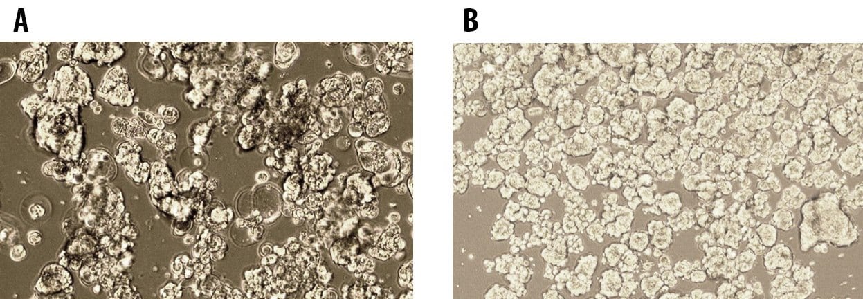
3D Tumorsphere formation of MCF7

Figure 1.3D Tumorsphere formation of MCF7 breast cancer and E006AA prostate cancer cells at Passage 5 cultured in 3dGRO™ Spheroid Medium.

3D Spheroid Formation Plates

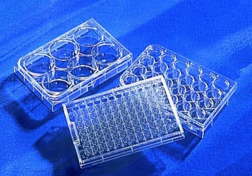
Corning® Costar® Ultra-Low attachment multi-well plates are designed to inhibit cell attachment to efficiently form 3D spheroids, including cancer stem cells. The surface is coated with a highly hydrophilic and neutrally charged coating that binds to polystyrene forcing seeded cells into a suspended floating state. The coating is stable, nontoxic, biologically inert and non-degradable.

3D Spheroid Formation Plates

Cancer Stem Cell Markers

Small populations of stem-like cells have been isolated from most leukemias as well as from many solid tumors such as brain glioblastomas, medulloblastomas, breast, cervical, colorectal, gastrointestinal, hepatocellular, lung, pancreatic, prostate, and skin carcinomas8. Cancer stem cells display the same cell surface markers as their normal counterparts, but demonstrate uncontrolled proliferation, perhaps due to a reduced responsiveness to negative growth regulators or to the loss of contact inhibition and gap junction intercellular communication (GJIC).

Table 1.Cancer Stem Cell Markers
Table 2.Cancer Stem Cell Antibodies

Cancer Stem Cell ALDH Detection

CSCs are unique as they express elevated levels of aldehyde dehydrogenase (ALDH) 9. Historically, the ALDEFLUOR™ assay has been used to measure ALDH levels and has been influential to study rare CSCs populations. The ALDEFLUOR assay emits in the green region of the electromagnetic spectrum (512 nm). Thus, the reagent cannot be simultaneously utilized in cells or mice expressing green-fluorescent proteins emitting in the green fluorescent spectrum. AldeRed is a red-shifted fluorescent substrate for ALDH used for labelling viable ALDH positive cells leaving the green emission channel available for further experimentation10. The AldeRed ALDH Detection Assay addresses current limitations of the ALDEFLUOR assay.

AldeRed Features and Benefits:

  • Red-shifted assay leaves green channel available for further experimentation
  • Live stem cell identification enables flow sorting of rare cell populations
  • Rapid enzymatic assay protocol
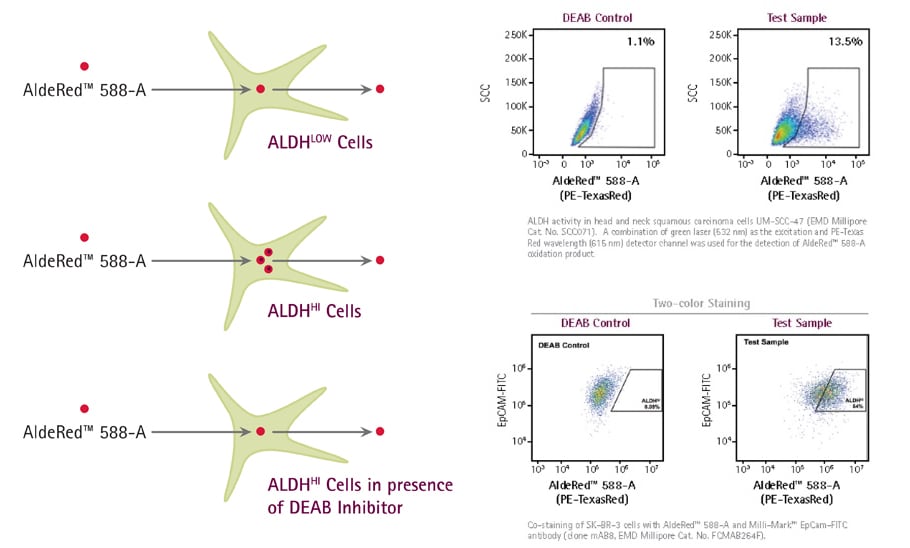
AldeRed 588-A Deab Control

Figure 2.AldeRed 588-A is a fluorescent and non-toxic ALDH substrate that diffuses freely into live intact and viable cells, but remains trapped inside the cells once converted by ALDH into the corresponding acid.

Materials
Loading

References

1.
Kreso A, Dick J. 2014. Evolution of the Cancer Stem Cell Model. Cell Stem Cell. 14(3):275-291. https://doi.org/10.1016/j.stem.2014.02.006
2.
Adams P, Jasper H, Rudolph K. 2015. Aging-Induced Stem Cell Mutations as Drivers for Disease and Cancer. Cell Stem Cell. 16(6):601-612. https://doi.org/10.1016/j.stem.2015.05.002
3.
Vinogradov S, Wei X. 2012. Cancer stem cells and drug resistance: the potential of nanomedicine. Nanomedicine. 7(4):597-615. https://doi.org/10.2217/nnm.12.22
4.
Karamboulas C, Ailles L. 2013. Developmental signaling pathways in cancer stem cells of solid tumors. Biochimica et Biophysica Acta (BBA) - General Subjects. 1830(2):2481-2495. https://doi.org/10.1016/j.bbagen.2012.11.008
5.
Plaks V, Kong N, Werb Z. 2015. The Cancer Stem Cell Niche: How Essential Is the Niche in Regulating Stemness of Tumor Cells?. Cell Stem Cell. 16(3):225-238. https://doi.org/10.1016/j.stem.2015.02.015
6.
Vidal SJ, Rodriguez-Bravo V, Galsky M, Cordon-Cardo C, Domingo-Domenech J. 2014. Targeting cancer stem cells to suppress acquired chemotherapy resistance. Oncogene. 33(36):4451-4463. https://doi.org/10.1038/onc.2013.411
7.
Wong DJ, Segal E, Chang HY. 2008. Stemness, cancer, and cancer stem cells. Cell Cycle. 7(23):3622-3624. https://doi.org/10.4161/cc.7.23.7104
8.
Nguyen LV, Vanner R, Dirks P, Eaves CJ. 2012. Cancer stem cells: an evolving concept. Nat Rev Cancer. 12(2):133-143. https://doi.org/10.1038/nrc3184
9.
Marcato P, Dean CA, Giacomantonio CA, Lee PW. 2011. Aldehyde dehydrogenase: Its role as a cancer stem cell marker comes down to the specific isoform. Cell Cycle. 10(9):1378-1384. https://doi.org/10.4161/cc.10.9.15486
10.
Minn I, Wang H, Mease RC, Byun Y, Yang X, Wang J, Leach SD, Pomper MG. 2014. A red-shifted fluorescent substrate for aldehyde dehydrogenase. Nat Commun. 5(1): https://doi.org/10.1038/ncomms4662
Sign In To Continue

To continue reading please sign in or create an account.

Don't Have An Account?