Patient-Derived Organoid and Immune Cell Biobanks for Immuno-Oncology Applications
Recent advances in cancer immunotherapy have had a positive impact on the life expectancy of patients with liquid cancers, whereas solid tumors remain an open challenge for immunotherapeutic development. The lack of clinically predictive biomarkers coupled with the poor translatability from conventional 2D cancer models represents a major hurdle for preclinical development.
Patient-derived organoids (PDOs) generated from healthy and malignant tissues recapitulate the complex characteristics of the original parental tissue, including molecular heterogeneity and morphological and functional traits. Importantly, they preserve tumor-specific antigens that are conventionally lost in standard in vitro models, therefore representing an excellent system to investigate efficacy, target engagement, and mechanism of action, and to stratify a patient population based on tumor molecular features.
In this article, we describe the development of PDO and immune cell biobanks relevant for testing immuno-oncology agents, as well as several co-culture assays.
Section overview
- Patient-derived organoids and drug discovery
- Immuno-oncology PDO biobank
- PDOs in immuno-oncology: Case studies
- Exploiting γδTCRS to target hematological tumors
- Harnessing PDO screens to expand GDT002 indications to solid tumors
- I-O biobanks support immunotherapeutic development
- Frequently asked questions about I-O biobanks
Patient-derived organoids and drug discovery
The current drug development paradigm has proven over the years to be an inefficient, unpredictable, and expensive process, with a high attrition rate of new compounds in the clinic. This is partly due to a lack of clinically relevant preclinical models that can be used as patient avatars to test treatment responses before entering clinical trials. Drug development can be improved by replacing standard preclinical models with patient-derived organoids (PDOs) that hold a high predictive value of patient response in the clinic. PDOs’ predictive value of patient response in the clinic has been extensively demonstrated by multiple independent clinical validation studies published in high-impact peer-reviewed papers1-3 and led in 2015 to treating the first cystic fibrosis patient4 with ultra-rare mutations based on organoid data. More recently, a proof-of-concept study5 was published demonstrating the feasibility of progressing a new oncology clinical candidate to clinical trials using organoid screening.
PDOs – or HUB Organoids® – are adult stem cell-derived organoids that capture patient and tumor heterogeneity and mimic patient response to treatment. These organoid models are physiologically relevant and genetically and phenotypically stable.
The HUB Organoid Technology allows for the development of PDO models from virtually any epithelial organ. Living biobanks that represent various human diseases can be generated, which undergo strict quality control and characterization to ensure they preserve original patient features over multiple passages and after cryopreservation. This allows researchers to recapitulate the diversity of the patient population in the lab and to conduct preclinical studies that enable patient stratification and the identification of biomarkers of response – something which is still lacking for most immunotherapies.
Immuno-oncology PDO biobank
Currently approved cell therapies which have shown success in hematological malignancies were developed to target linear specific markers. However, this approach has failed in solid tumors due to the lack of tumor-associated antigens (TAAs) that can be targeted while sparing normal tissue. When TAAs have been identified in patient tumors, standard preclinical models have failed to recapitulate TAA expression thus limiting therapeutic development. Interestingly to immuno-oncology (I–O) applications, PDOs preserve patient heterogeneity and specific TAAs, which is key for developing cell therapies for solid tumors.
Co-cultures of organoids can be set up with various immune cell types, either in an allogenic or autologous setting to investigate immune-therapeutics targeting the tumor, the immune microenvironment, or both. Thanks to HUB Organoid Technology’s unique culturing conditions, PDOs can be established from both normal epithelial and cancer lesions, in some cases from the same patient, thus allowing testing for off-target/off-tumor effects by comparing normal and tumor responses.
To adapt to the growing demand for patient-relevant models for immuno-oncology, HUB has focused on developing I–O biobanks, with protocols to isolate and expand not only tumor cells from resected tissue but also other cell types such as fibroblasts and T cells to establish autologous and non-autologous systems. These complex biobanks will enable us to dissect the role of different components of the tumor microenvironment in treatment response.
PDOs in immuno-oncology: Case studies
Figure 1 shows morphology read out and T cell activation following allogenic co-culture of engineered T cells and organoids. Tumor and normal organoids were placed in screening wells in combination with engineered T cells designed to identify and kill tumor organoids based on antigen recognition. After co-culture for 3 days, bright field images confirmed tumor organoid killing, whereas no significant morphological changes were detected in the normal organoid culture.
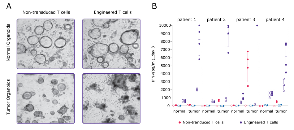
Figure 1.A. PDO and engineered T cell co-cultures. B. Cytokine analysis to evaluate tumor organoid killing and T cell activation.
IFNу measurement confirmed engineered T cells activation in the presence of tumor organoids but not normal organoids.
Additional readouts were developed to offer alternative options to explore the activity of these cellular products. A typical chimeric antigen receptor (CAR)-T testing workflow (Figure 2) first involves organoid model selection based on CAR-T target antigen expression by tumor cells. Subsequently, a co-culture is established, and CAR-T cell activation is detected by ELISA IFNg measurement. In this example, an imaging-based readout (caspase 3/7 signal) is used to measure tumor organoid killing over time by CAR-T cells. Alternative readouts are also under development to measure a larger spectrum of activities depending on the compound mode of action.
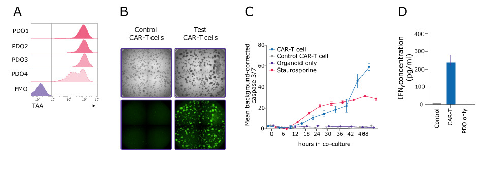
Figure 2.CAR-T cell testing workflow. Imaging-based readouts coupled with cytokine analysis provide an overview of CAR-T efficacy.
Autologous PDO and T cell co-cultures have been adopted in this sample study to assess the efficacy and tumor specificity of bispecific antibodies (Figure 3). In this example, T cells were isolated and expanded from patient peripheral blood mononuclear cells (PBMCs), and PDOs were selected based on target tumor antigen expression, with tumor PDOs expressing high levels of target antigen and matched normal PDOs expressing lower antigen levels, similarly to corresponding patients. No killing of normal organoids was detected by caspase 3/7 signal, even in the presence of high-dose T cell bispecific antibody, whereas dose-response curves display a correlation between increasing therapeutic dose and tumor PDO killing.
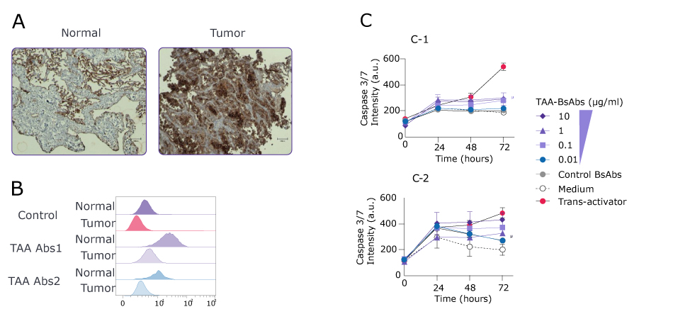
Figure 3.PDO and PBMC-derived T cell co-cultures to assess the efficacy of bispecific antibodies. Sample study workflow for an anti-CD3/anti-TAA bispecific (BsAbs).
Exploiting γδTCRS to target hematological tumors
γδ-T cells are a subpopulation of T cells, either tissue-resident or circulating, with unique features that allow them to recognize stress-related molecules on cells that experience some level of alteration, including malignant transformation. Recent publications have shown that the presence of γδ-T cells within tumors is associated with a better patient prognosis, suggesting their general role in counteracting tumor progression. At the molecular level, γδ-T cells target cancer cells by recognizing surface antigens using γδ-T cell receptors (γδTCRs) in a human leukocyte antigen (HLA)-independent manner. This offers the possibility of using γδTCRs in a broad spectrum of patients.
Gadeta, a biotechnology company founded in 2015 and based in Utrecht, is focused on harnessing the unique capacity of γδTCR to target tumors for the development of first-in-class cell therapies. Their innovative technology led to the development of GDT002, a first-in class cell therapy targeting CD277, a tumor cell antigen presented on the surface of cancer cells with altered bisphosphonates metabolisms (Figure 4).
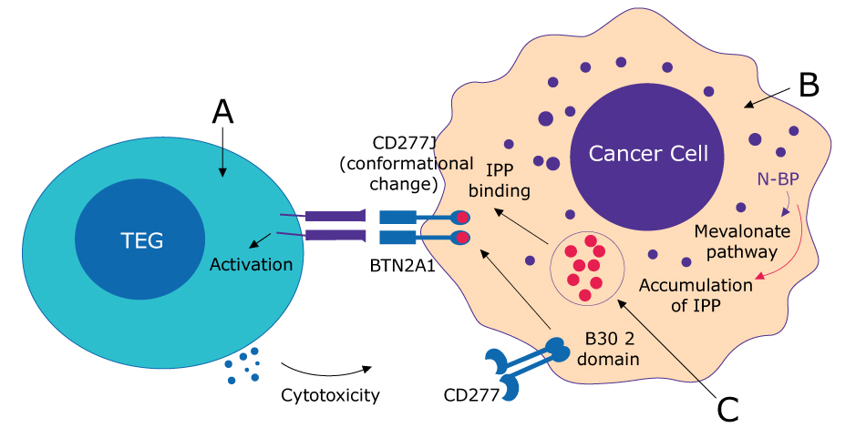
Figure 4.GDT002 mechanism of action. A. αβ T cells transduced with clonal у9б2 TCR construct; B. GDT002 recognizes a metabolically altered surface target; C. Malignant transformation can alter metabolism
GDT002 has broad tumor reactivity but does not cause cross-activity to healthy tissue from different organs. It has now been tested in a multicenter Phase 1/2 clinical trial evaluating safety, tolerability, and preliminary efficacy in patients with relapsed or refractory multiple myeloma.
Harnessing PDO screens to expand GDT002 indications to solid tumors
Given the positive results obtained with GDT002 in hematological cancer, Gadeta was interested in expanding the application of the drug to solid tumors and selected a PDOs basket trial as a patient-relevant platform to obtain preclinical data for their IND package submission. GDT002 showed a broad reactivity against tumor PDOs, with a remarkable 90% reactivity against ovarian PDOs (Figure 5). Therefore, ovarian cancer was prioritized as the chosen indication for the first clinical trial to assess the safety of GDT002 in solid tumors.
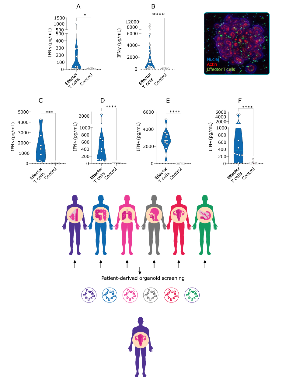
Figure 5.GDT002 shows broad reactivity against tumor PDOs. Effector T cells (GDT002) and PDOs co-culture to evaluate anti-tumor activity on a panel of different cancer types.
I-O biobanks support immunotherapeutic development
We show that autologous PDO and immune cell biobanks can be co-cultured for testing a variety of T cell targeting therapies. Building these patient-relevant biobanks supports immunotherapeutic development by providing a scalable and physiologically relevant system that recapitulates patient heterogeneity and preserves key TAA to investigate immune cell activation and tumor cell killing downstream of target engagement. PDOs co-cultures with autologous or allogenic immune cells allow to test the efficacy and off-target toxicities in parallel, thus providing a comprehensive profile of the therapeutic efficiency of a new agent before moving with confidence to patient trials.
Frequently asked questions about I-O biobanks
How large are your I–O biobanks?
Our immuno-biobanks began recently. In the context of colorectal (CRC) models, we have a dozen models from which we have isolated tumor-infiltrating lymphocytes (TILs), and in non-small cell lung cancer, we have around five models. In a subset of these CRC immuno-oncology (I–O) models, we have isolated cancer-associated fibroblasts (CAFs). We are also interested in indications like bladder and head and neck cancers, where we see a lot of interest in I–O. Patient-derived biobanks are not limited to particular indications – we can develop specific biobanks depending on need.
How do you identify the ligands of γδTCRS?
Our approach to identifying new tumor-reactive γδTCRs is based on functional assays: initially, we want to ensure that these γδTCRs are tumor-specific and not recognizing any healthy cells. Then, it is important to identify the ligand on tumor cells. We normally do this through two approaches. One is functional genetic screening. The second is based on a more classic biochemical approach. The identification of γδTCR ligands has been and still is a challenge in the field: we think that a combination of these two approaches is the right way to tackle this aspect.
How can I work with HUB to develop I–O compounds?
We have different service offerings. I would advise anyone interested in our services to contact our business development (BD) team and explain the mechanism of action and the area in which your compound is developed. Our team can offer different solutions and approaches, either from existing assets or ones that require further development. Our strong scientific team can address any challenge and offer a solution.
Is there an option to validate target expression in both the patient material and in the organoids to control for differential expression between in vivo and ex vivo situations?
One challenge we face is that when we receive the tissue to establish the organoid, we know that there are interests for different tumor antigens but performing a complete analysis on the tissue to make comparisons is difficult. At a basic level, we must use immunohistochemistry, which is not quantifiable but can confirm that the tissue expresses a specific antigen. Then, we can confirm it is also expressed on organoids with flow analysis. If there is a particular application in which this is an important question, we could run a project in which we get new tissue for PDO generation. While establishing the organoids, we can characterize the original tissue using flow analysis. It is outside our standard activities, but we can address this.
How do you expand the tumor PDOs without losing the original phenotype and genotype, and how many passages can be maintained?
The culture conditions for expanding organoids were first established by culturing healthy cells to ensure we could expand stem cells, proliferative cells, and other cell types. We use the same principles to establish the tumor model. We believe that because there is no high selection pressure, we can maintain the original tumor heterogeneity in organoids.
When the first tumor biobanks were generated, original tissue and derived organoids were sequenced. In most cases, there was more than 80% overlap between driver mutations detected in original tissue and organoids. Of course, tumors are, by definition, genetically unstable, but we know that culture conditions do not drive this genetic instability. When working with tumor-derived organoids, we recommend expanding them for a maximum of 5–6 passages, as an average and depending on the model. We do not recommend expanding a tumor model for a year because it can vary too much from the original tumor. However, 2–3 months of expansion and cryopreservation can allow us to preserve the genetic landscape that the original tumor contained.
Based on the results of assessing your two treatments in organoid models, do you have plans to use organoids for future projects?
To characterize γδTCR tumor reactivity we proceed with a step-by-step approach. We start with tumor cell lines in vitro for the first layer of investigation. As soon as possible, we move selected γδTCRs into more clinically relevant models. For this, our collaboration with HUB is beneficial. Most of our γδTCRs can recognize broadly different tumor types, so we want to narrow down the tumor types that can be selected to be used in an initial clinical trial. We also would like to understand whether there is a genetic setting that is preferentially targeted. For both of these aspects the use of organoid models provides valuable information and will certainly be included in our strategy.
How do you know the reactivity is tumor-specific?
Our first layer of characterization of γδTCRs is a screen for lack of cross-reactivity against a large set of healthy primary cells to ensure there is not recognition of healthy vital tissues. Then, we test a set of tumor models, including organoids, to show that tumor cells are killed. In the next steps, we use mixed toxicology and pharmacology models with primary material from patients, to show specific killing of tumor cells, sparing the normal tissues in the samples.
In the allogeneic setting, could this T cell reactivity be due to HLA mismatch?
It is true that in most of the models there is a potential allogeneic reaction due to human leukocyte antigen mismatch. To control for this, we use either untransduced T cells, or our recently developed set of control TCRs that are engineered to not recognize tumor tissues. These two controls can be used for measuring the background level of possible allogenic activity.
Is there an imaging platform that can be used for organoids derived from patients and grown without dissociation into single cells, but as small tissue chunks?
When we perform our screens, the organoids are not single cells but structures. The average size of these organoids in our screening assays is ~50–70 μm in diameter. We use confocal microscopy in a high throughput format for imaging. As we are developing the technology, we are also working on selecting the best imaging platform for our data. There are several publications from different labs that can achieve good quality imaging data from screens performed on organoids. We do not see imaging as a limitation.
For co-culturing methods such as organoids with T cells, can I use non-activated T cells?
Yes, this is possible. In our co-culture assays, we have a control of activated T cells, but we have set up co-cultures where, for example, we wanted to evaluate tumor reactivity on TILs isolated from tumors without pre-activating the cells. We have seen a response with this, so it is possible to see cell activity.
What do you see as the most exciting application for organoids, now and in the future?
The developments we are working on in I–O and inflammatory diseases by combining organoids with other cell types is an exciting area. Our organization is also putting effort into validating the predictive value of organoids to show that organoids can predict patient response. A solution in the industry could be to run ‘avatar’ clinical trials with organoids before moving into patients to identify the patient populations likely to succeed in clinical studies. We believe that our technology will significantly contribute to this, and we are putting great effort into this.
Looking to use HUB Organoids® for your drug development program? Speak to an expert now!
Related products