Live Cell Imaging Reagents
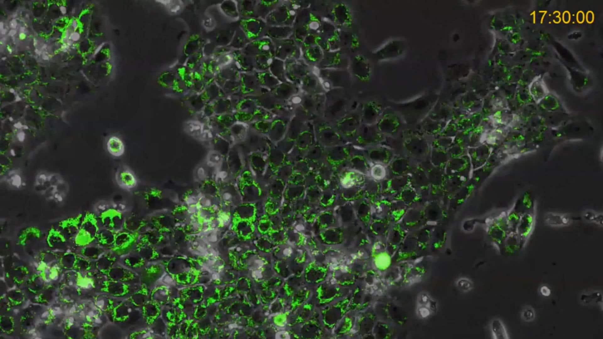
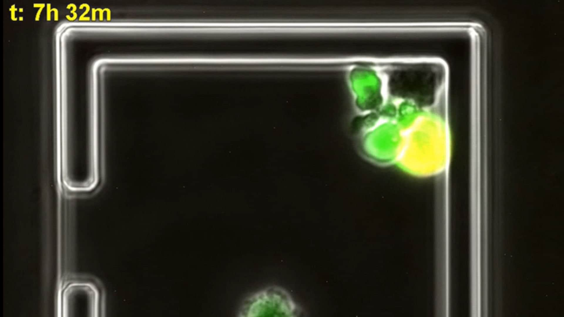
Live cell imaging in real time allows scientists to monitor dynamic cellular events that otherwise escape observation using traditional endpoint techniques like fixed imaging and PCR that capture only a static moment in time. Our portfolio of live cell imaging reagents includes novel fluorescent cell labeling technologies for cell tracking, fluorescent lentiviral biosensors, traditional fluorescent dyes and probes to analyze cellular events in real-time. These reagents are optimal for capturing dynamic cellular events with real-time incubation and imaging systems like the CellASIC® ONIX2 Microfluidic live cell imaging system, but can also be used like traditional fluorescent cell culture reagents with standard or confocal fluorescence microscopy.
Featured product descriptions continue below product table, including for PKH and CellVue® Fluorescent Cell Linker Labeling, LentiBrite™ Fluorescent Lentiviral Biosensors, BioTracker® Live Cell Fluorescent Dyes and Stains, and more.
Products
PKH and CellVue® Fluorescent Cell Linker Labeling
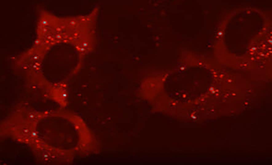
PKH and CellVue® Fluorescent Cell Linker Kits provide fluorescent membrane labeling of live cells for long-term live cell imaging experiments.
Key attributes of PKH and CellVue® dyes include:
- Stable for up to 100 days on live cells in vivo
- Safe for eukaryotic cells, bacterial cells, and parasites
- No significant dye leakage or cell-to-cell transfer
- Uniform and reproducible signal
- Spectrally different dyes allow multicolor analysis
- Compatible with other fluorescent labels
- Cited in over 7,000 publications
LentiBrite™ Fluorescent Lentiviral Biosensors
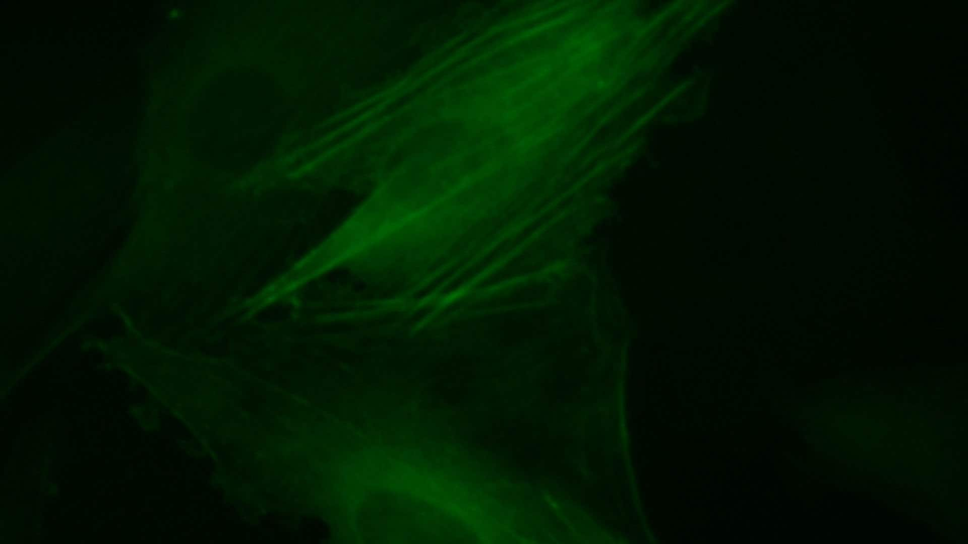
LentiBrite™ Fluorescent Lentiviral Biosensors are a suite of pre-packaged lentiviral particles that code for fluorescent proteins associated with autophagy, apoptosis, and cell structure for insights from these processes in live cells.
Lentiviral Biosensors feature:
- Pre-packaged, ready-to-use format, tagged with monomeric GFP & RFP
- Minimum titer (≥3 x 10⁸ IFU/mL) per vial
- Superior transfection efficiency to chemical-based methods
- For dividing, non-dividing, and hard-to-transfect cells like primary cells or stem cells
- Non-disruptive of cellular function
BioTracker® Live Cell Fluorescent Dyes and Stains
BioTracker® Live Cell dyes are cell-permeable dyes for live cell applications such as organelle labeling, apoptosis detection, cell viability and cell health analysis, hypoxia monitoring, reactive oxygen species tracking, calcium indicator function, and neural and stem cell (SCR150, SCT029, SCT046) cultures.
LuminiCell Tracker™ Fluorescent Nanoparticles
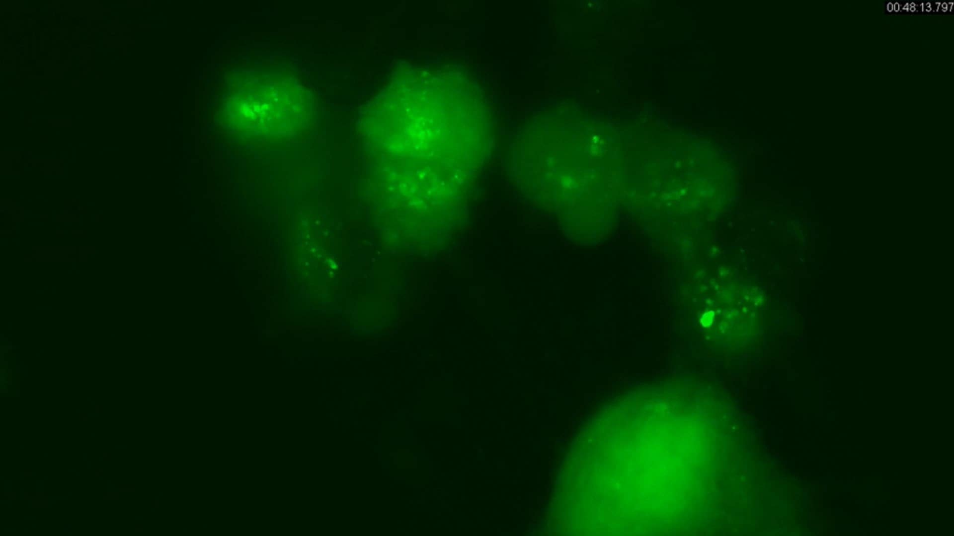
LuminiCell Tracker™ fluorescent nanoparticles provide an effective, simple, and nontoxic reagent for long-term cell tracking experiments. Based on aggregation-induced emission (AIE) technology, these cell tracking dyes have been encapsulated into biocompatible, cell-permeable, fluorescent nanoparticles (AIE Dot) that provide enhanced photostability, ultra-bright fluorescence, and low cellular toxicity for bioimaging applications over extended periods of time.
- Brighter: 10X brighter than many alternative cell labeling technologies
- Photostable: 3X fluorescence longevity, without signal quenching
- Biocompatible: Nontoxic organic nanoparticles developed for biological applications
- Rapid Protocol: Easy-to-follow protocol labels cells in four hours or less
Available in multiple fluorescence emissions to optimize multiplexing, our newest AIE cell tracker (SCT017) is in the essential NIR (near-infrared) range to enhance application versatility and expand multiplex potential.
Related Resources
- Article: Live Cell Imaging of β-Actin Cytoskeleton Proteins using LentiBrite™ Fluorescent Biosensors
Learn how these pre-packaged lentiviral particles facilitate visualization of autophagy, apoptosis, and cell structure in live cell analysis.
- Article: Long-Term Live Cell Tracking of Cancer and Stem Cells Using a Biocompatible Fluorescent Nanoparticle Based Upon Aggregation Induced Emission (AIE Dot) Nanotechnology
Biocompatible organic fluorescent nanoparticles (AIE Dots) that can be used to label cells and vasculature for long-term live cell tracking and tracing experiments.
- Article: Lipophilic Dye Kits for Cell Tracking
Lipophilic cell tracking dyes enable cancer biologists to track tumor and immune cell functions both in vitro and in vivo. Read the article to choose a right membrane dye kit for cell tracking and proliferation monitoring.
- Article: Exosome Labeling Protocol with PKH Lipophilic Dyes
PKH dyes label exosomes for tracking experiments in vitro and in vivo, detailed protocol provided.
- Article: Fluorescent Live Cell Imaging of Cytoskeleton Structure Proteins using LentiBrite™ Lentiviral Biosensors
High titer lentiviral particles including beta-actin, alpha-tubulin and vimentin used for live cell analysis of cytoskeleton structure proteins.
- Article: Fluorescent Live Cell Imaging of LC3 Mediated Autophagy using LentiBrite™ Lentiviral Biosensors
High titer lentiviral particles for LC3 variants used for live cell analysis of cellular autophagy.
- Article: Cancer Stem Cell Detection with AldeRed™ 588-A
AldeRed™ 588-A detects ALDH activity in cancer stem cells, aiding in cancer research.
- Article: PKH and CellVue® Claret Fluorescent Cell Linker Dyes
PKH and CellVue® Fluorescent Cell Linker Kits provide fluorescent labeling of live cells over an extended period of time, with no apparent toxic effects.
- Article: Live Cell Fluorescent Organelle Dyes and Stains
BioTracker fluorescent organelle stains and dyes bring experiments to life with multiple color options.
- Article: Dynamic Live Cell Imaging of Caspase Mediated Apoptosis and Cellular Hypoxia in Lymphocytes and Cancer Cells Using the CellASIC® ONIX2 Microfluidic Platform
Article on how the CellASIC® ONIX2 microfluidic system can be used to analyze caspase-3 mediated apoptosis/cell death and cellular hypoxia in live immune and cancer cell lines.
To continue reading please sign in or create an account.
Don't Have An Account?