TrueGel3D™ Features and Benefits
How to use the TrueGel3D™ system
Selecting the right TrueGel3D™ kit for your application
Request Information
Introduction
TrueGel3D™ is a biochemically defined hydrogel formed by mixing polymers with crosslinkers. Compared to other hydrogels based on a biological extract from animal cells (e.g. mouse EHS tumor cells), TrueGel3D™ does not contain any products of animal origin that could interfere with or contaminate experiments. Moreover, components that are included in TrueGel3D™ preserve viability and allow critical features of the natural extracellular matrix (ECM) to be mimicked. They replicate native cellular environments similar to those of tissues by supporting cell adhesion and migration. TrueGel3D™ technology provides mechanical and biochemical cues to investigate both morphological and physiological properties of cells in the 3D environment. Finally, these hydrogels allow encapsulation of cells for growth in a 3D environment that more closely mimics native tissue environments when compared with traditional 2D cell culture conditions.
TrueGel3D™ Features and Benefits
- TrueGel3D™ works with a multitude of cells (MDCK, epithelial, fibroblast, cancer, primary including lymphocytes, stromal and embryonic heart muscle cells).
- TrueGel3D™ basic components (dextran, PVA, PEG) form a basic hydrogel scaffold that does not interfere with cells
- Gels are transparent, enabling cell imaging capabilities
- Multiple TrueGel3D™ Kits available with different gel speeds, supporting a variety of applications (summarized in Table 2)
- Non-toxic cell recovery option
Most importantly, TrueGel3D™ has a defined composition, unlike basement membranes extracts derived from animal cells where a mix of ECM components and growth factors can interfere with the cells because it creates a non-native environment.
Protocols and Support
- Protocol 1: Preparation of TrueGel3D™ hydrogel, CD cell-degradable crosslinker and RGD peptide (TRUE1)
- Protocol 2: Preparation of Fast hydrogels (True2-5)
- Protocol 3: Preparation of Slow hydrogels (True6-9)
- Advanced protocol for hydrogel optimization
- Application Notes
- High-Throughput 3D Cell Culture Using TrueGel3D®
- Cyst formation of MDCK cells application note
- Fibroblast spreading
- Co-culture of tumor and stromal cells
How does TrueGel3D™ work?
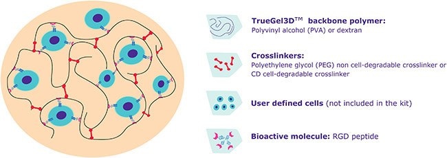
The TrueGel3D™ technology is a four component system consisting of:
- TrueGel3D™ polymer: either polyvinyl alcohol (PVA) or dextran
- Crosslinkers: two different thiol-functionalized crosslinkers (polyethylene glycol (PEG) or cyclodextrin (CD)) that provide a chemical bond to connect polymer chains and form the hydrogel
- Cell-interactive components: when cell adhesion to the matrix is required, peptides including adhesion sequences like TrueGel3D™ Arg-Gly-Asp (RGD) integrin adhesion peptide can be added to the mix. Other bioactive components like extracellular matrix proteins (e.g. fibronectin, laminin), peptides, heparan sulfate or growth factors (not included in kits) can also be added.
- User-defined cells (not included in kits)

Figure 1.TrueGel3D™ technology

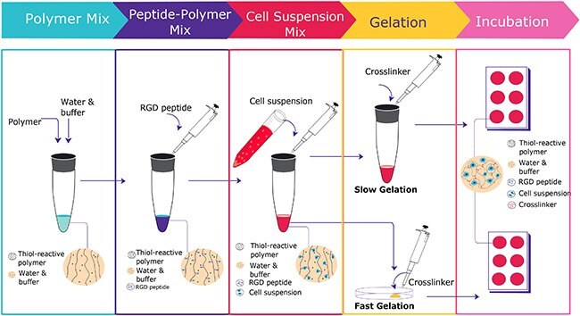
Figure 2.TrueGel3D™ protocol flowchart
- Will a predefined system work for me, or do I need to customize my environment?
For many applications like spheroid formation or co-culture models, you can use the pre-configured TrueGel3D™ hydrogel, CD cell-degradable crosslinker and RGD peptide (TRUE1-1KT). This hydrogel contains a defined concentration of cell adhesion sites, allows cell spreading and cell migration, and can be degraded for cell recovery by using TrueGel3D™ enzymatic cell recovery solution (TRUEENZ-500UL). This gel offers a moderate stiffness and a complete gel formation in around 20 minutes.
If you need to optimize your gel environment (e.g. change in stiffness, adhesion molecules, ECM proteins, etc.), other TrueGel3D™ kits are available and are listed in Table 1.
- What gelation rate is optimal for my application?
*During this time, the mix with all components remains liquid; after that, it begins to form a gel and can no longer be pipetted.
**For a more detailed view of recommended gels by application, please refer to Table 2.
The moderate gelation time is only available for pre-configured TrueGel3D™ hydrogel, CD cell-degradable crosslinker and RGD peptide (Cat. No.TRUE1-1KT). Fast and slow gelation kits are listed in Table 1.
- Do I need to add cell-interactive components?
For some applications like cell or tissue immobilization for microscopy, you can use TruGel3D™ kits without any additional active components. TrueGel3D™ RDG peptide is required for cell adhesion and spreading (presence of the CD degradable crosslinker is also necessary for cell spreading). You can also add other cellular components like growth factors*, heparin or ECM proteins (e.g. fibronectin or collagen) in your hydrogel.
*Be aware that some components may diffuse outside the gel, so should instead be added to the cell culture medium.
- Is cell recovery needed for further analysis or culture?
Cells can be recovered from dextran based TrueGel3D™ backbones using TrueGel3D™ enzymatic cell recovery solution (see advanced protocols for more details). Cells can’t be recovered from PVA based TrueGel3D™ backbone.
- Does my application require a cell-degradable hydrogel?
If you want to study cell spreading or motility, a cell-degradable environment is required. We offer a selection of TrueGel3D™ kits containing a CD cell-degradable crosslinker, which includes matrix metalloprotease (MMP) cleavage site peptide sequences*. By degrading the crosslinker, cells can spread inside the hydrogel (in the presence of a cell-adhesive ligand like RGD integrin adhesion peptide).
*Not all MMPs have been shown to cleave the CD link. CD-Link contains the sequence Pro-Leu-Gly-Leu-Trp-Ala, which is known to be cleaved at least by MMP1, MMP3, MMP7 and MMP9.
*TrueGel3D™ RGD adhesion peptide (provided as separate component) needs to be added
Chemistry Overview
Polymers and crosslinkers with differential properties provide options for gel requirements
TrueGel3D™ hydrogel systems employ one of two types of polymers: a synthetic, non-degradable polyvinyl alcohol (PVA) or an enzymatically-degradable dextran. Both polymers are functionalized either with fast or slow thiol-reactive groups.
- Fast thiol-reactive groups on the polymer backbone react rapidly with crosslinkers to form hydrogels within a few minutes.
- Slow thiol-reactive groups on the polymer backbone react at a slower rate and form hydrogels more slowly.
Polymers (PVA and dextran) are inert and do not adhere to cells. However, they can be conjugated with bioactive components such as adhesion peptides or combined with extracellular matrix proteins to allow cell adhesion and mimic relevant physiological conditions.
Crosslinkers in TrueGel3D™ are also of two types. The PEG non-cell-degradable crosslinker is made up of polyethylene glycol with thiol (SH) groups at each end. The CD cell-degradable crosslinker is similar to PEG non-cell-degradable crosslinker, but includes peptidic sequences, creating cleavage sites for matrix metalloproteases (MMPs). Gels with CD cell-degradable crosslinker allow cells to spread and migrate by cleaving conjugated peptide if cell adhesion molecules are also a component of the gel.
Variables affecting the gelation rate:
Gel formation occurs best at equimolar concentration (starting from 1.8 mmol/L) of thiol (crosslinker) and thiol-reactive groups (polymer). However, a stiffer gel can be formed with a higher concentration of thiol and thiol-reactive groups.
In addition, the reaction rate is also controlled by hydrogen ion concentration (pH). At a lower pH (acidic environment) the deprotonated thiolate anions react slowly with thiol-reactive groups and slow down the network formation between polymer and crosslinker. Thus, buffers in the kits (pH 5.5 and 7.2) can be mixed to generate an intermediate pH to aid in optimization of gelation time. Both buffers include phenol red to facilitate pH monitoring.
Video:Beating Cardiac Muscle Cells Cultured in TrueGel3D™ Synthetic Hydrogels
A suspension of freshly prepared cells from embryonic chick heart tissue was embedded in a Dextran-based hydrogel modified with RGD Peptide and crosslinked with matrix metalloprotease-cleavable crosslinker (TRUE1). Aggregated heart muscle cells are surrounded by fibroblasts. Heart muscle cells courtesy of Dr. Udo Kraushaar (NMI Reutlingen, Electrophysiology Group, Germany).
Materials
To continue reading please sign in or create an account.
Don't Have An Account?