Hematopoietic Stem & Immune Cell Culture
- Hematopoietic Stem Cells
- Human Monocytes
- Human Mononuclear Cells (PBMCs)
- Human Macrophages
- Human Dendritic Cells
- Stromal Cells for T-Cell Differentiation
Hematopoietic stem cells (HSCs) give rise to all blood cell types including myeloid and lymphoid lineages. Myeloid cells include monocytes, macrophages, neutrophils, basophils, eosinophils, erythrocytes, dendritic cells, and megakaryocytes or platelets. Lymphoid cells include T cells, B cells, and natural killer cells. HSCs are found in the bone marrow of adults, umbilical cord blood, placenta, and mobilized peripheral blood. Recent advances in hematopoietic stem cell biology have resulted in the use of HSC transplants in the treatment of cancers and other immune system disorders. We offer a broad range of tools, cells and optimized serum-free expansion media to culture CD34+ human hematopoietic stem cells and other human immune cell types including CD14+ monocytes, mononuclear cells (PBMCs), macrophages, dendritic cells and T-cell lymphocytes.
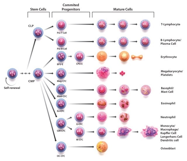
Figure 1. Stages of hematopoiesis.Through the process of hematopoiesis, all cellular blood components are derived from hematopoietic stem cells.
Hematopoietic Stem Cells
CD34+ Hematopoietic Progenitor Cells
CD34 is a marker of human HSCs. All colony-forming activity of human bone marrow (BM) cells is found in the CD34+ progenitor cell fraction. Clinical transplantation studies that used enriched CD34+ BM cells indicated the presence of HSC with long-term BM reconstitution ability within this fraction. We offer cryopreserved CD34+ hematopoietic progenitor cells isolated from the cord blood of healthy human donors. The CD34+ progenitor cells contain two main cellular subpopulations, hematopoietic and endothelial progenitor cells. Therefore, CD34+ progenitor cells are suitable for a series of studies including directed differentiation into more committed types of blood cells and endothelial lineages.
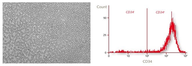
Figure 2.Morphology and characterization of human CD34+ hematopoietic progenitor cells by flow cytometry.
Hematopoietic Stem Cell Media
HSCs are able to expand in vivo to large numbers by virtue of their pronounced self-renewal abilities. The in vitro colony forming unit (CFU) assay has been used to study the proliferation and differentiation pattern of hematopoietic progenitors by their ability to form colonies in a semisolid agar medium. However, researchers face difficulties during in vitro expansion of undifferentiated HSCs, with increasing differentiation of the cells observed during the expansion phase in traditional serum-containing culture systems.
The Hematopoietic Progenitor Cell (HPC) Expansion Medium DXF has been designed for the maximum expansion of primitive HPCs in a fully defined, xeno-free culture environment. Due to the utilization of exclusively synthetic, recombinant or plant-sourced materials, this medium is defined and free of all animal-derived components.
Application Note: In Vitro Expansion of CD34+/CD38- Human Hematopoietic Progenitor Cells in a Serum-free and Xeno-free Stem Cell Culture Media.
Featured Product
Stemline® II Hematopoietic Stem Cell Media
Developed to promote the optimal expansion of human hematopoietic stem cells (HSC) from bone marrow, mobilized peripheral blood, and cord blood, Stemline® Hematopoietic Stem Cell Expansion Medium (S0192) demonstrates higher total nucleated cell (TNC) fold increases than other commercially available serum-free media formulations.
Features and Benefits
- Serum-free formulation
- Enhanced expansion from cord blood CD34+ cells
- Expands cells from all appropriate hematopoietic lineages in a colony-forming unit.
- Tested extensively in 7-day and 14-day growth assays.
- Produced in a GMP state-of-the-art facility with an available Device Master File (DMF).
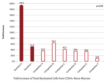
Figure 2. Stemline® demonstrates superior expansion of bone marrow hematopoietic stem cells (HSC).CD34+ HSCs were seeded into the wells of 24-well tissue culture plates. One milliliter of medium was added to each well with the appropriate cytokines to stimulate growth (100ng/mL each of TPO, SCF and G-CSF). Each condition was performed in triplicate and seeded with 10,000 cells per ml in each well. Cells were counted on day 14 and the fold increase was determined by cells final/cells initial. HSCs from bone marrow cultured in Stemline® and Stemline® II demonstrated superior expansion to those grown in other serum-free HSC media.
Hematopoietic Cytokines and Growth Factors
Numerous immune related cytokines are involved in the regulation of hematopoiesis within a complex network of positive and negative regulators. Some cytokines have very narrow lineage specificities of their actions, while many others have rather broad and overlapping specificity ranges. Listed within this section are the cytokines and growth factors whose action appears to be the stimulation of or regulation of hematopoietic stem cell development:
Hematopoietic Stem Cell Markers
AldeRed™ ALDH Detection Assay
Hematopoietic stem cell express elevated levels of aldehyde dehydrogenase (ALDH). ALDH activity has been used as a marker for the quality of hematopoietic stem cell transplants. Historically, ALDH levels have been measured using live cell-permeable dyes that emit in the green region of the electromagnetic spectrum (512 nm). Thus, the reagent cannot be simultaneously utilized in cells or mice expressing green-fluorescent proteins emitting in the green fluorescent spectrum. The AldeRed™ ALDH Detection Assay is a red-shifted fluorescent substrate for ALDH used for labelling viable ALDH positive HSCs leaving the green emission channel available for further experimentation.
Human Monocytes
Monocytes are immature phagocytic cells circulating in the blood. Acting as antigen-presenting immune cells, they can phagocytize and degrade microbes and particulate matter. Our primary human CD14+ monocytes are isolated from fresh peripheral blood of a single healthy donor. After isolation of the ultra-pure mononuclear cell fraction by proprietary methods, the CD14+ Monocytes are isolated by positive selection with > 95% purity. The Monocyte Attachment Medium allows for the efficient adherence selection of monocytes from freshly isolated human mononuclear cells while maintaining optimal cell health.
Human Mononuclear Cells (PBMCs)
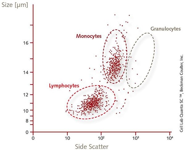
Mononuclear cells, including peripheral blood mononuclear cells (PBMCs), represent the lymphocyte and monocyte fraction isolated from umbilical cord blood or peripheral blood. They are prepared by optimized low-density gradient centrifugation, which effectively reduces the granulocyte content. Red blood cells are gently but thoroughly depleted by proprietary techniques instead of osmotic lysis. This results in ultra-pure mononuclear cells with superior viability and unchanged biological function. PBMCs can be used directly for experiments or as a starting material for the isolation of several cell types, e.g., hematopoietic and endothelial progenitors, monocytes, and lymphocyte subpopulations.
Human Macrophages
Macrophages are tissue-resident professional phagocytes and antigen-presenting cells (APC), which differentiate from circulating peripheral blood monocytes into either M1 or M2 phenotypes. The Macrophage Generation Media has been developed for the efficient generation of monocyte-derived macrophages (MDM) from freshly isolated human peripheral blood monocytes or directly from PBMCs as a starting material. The M1-Macrophage Generation Medium DXF contains GM-CSF and allows for the generation of M1-polarized macrophages. The M2-Macrophage Generation Medium DXF contains M-CSF and allows for the generation of M2-polarized macrophages. Both Macrophage DXF media are defined and xeno-free formulations and uses only exclusively synthetic, recombinant or plant-sourced materials, this medium is free of all animal-derived components.
Application Note: In Vitro Differentiation of Human PBMC Derived Monocytes into M1 or M2 Macrophages in a Serum-free and Xeno-free Cell Culture Media.
Human Dendritic Cells
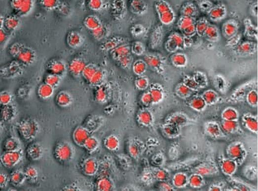
Dendritic cells are highly motile immune cells and are the most powerful antigen presenting cells (APCs) of the mammalian immune system. Dendritic cells are the only antigen-presenting cell capable of stimulating naive T cells, and hence they are pivotal in the generation of adaptive immunity. The Dendritic Cell (DC) Generation Medium has been developed for the easy and efficient generation of immature as well as fully mature myeloid Dendritic Cells from peripheral blood monocytes. DC Generation Medium is a defined and xeno-free formulation for use with freshly isolated cells. Due to the utilization of exclusively synthetic, recombinant or plant-sourced materials, this medium is free of all animal-derived components.
Application Note: In Vitro Differentiation of Human PBMC and CD34+ Derived Monocytes into Mature CD83+/CD14- Dendritic Cells.
Stromal Cells for T-Cell Differentiation
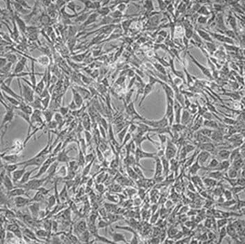
T lymphocytes develop in the thymus from bone-marrow derived hematopoietic stem (HSCs) and progenitor cells (HSPCs). In vitro, various model systems have been employed to mimic thymic function supportive of T-cell development. Recently, a new serum-free artificial thymic organoid (ATO) system was developed that supports highly efficient and reproducible in vitro differentiation and positive selection of human T-cells from multiple sources of HSPCs, including cord blood, bone marrow and peripheral blood CD34+ HSPCs. A key component of the ATO system is the MS5-DLL1 cell line which eliminates the need for primary thymic tissues. To form organoids, MS5-DLL1 stromal cells are aggregated with HSPCs by centrifugation and cultured on a Millicell® cell culture insert in defined serum-free media.
To continue reading please sign in or create an account.
Don't Have An Account?