Mesenchymal Stem Cell FAQs
Mesenchymal stem cells (MSCs) are defined as a self-renewing population of adherent multipotent progenitor cells with the capacity to differentiate into several mesenchymal cell lineages including bone, cartilage and adipose tissue. Recently, mesenchymal stem cells have become cells of increased interest due to their use in potential allogenic stem cell therapies.
What are the types of mesenchymal stem cells?
Where do mesenchymal stem cells come from?
They are majorly isolated from bone marrow. However, cells which display characteristics of mesenchymal stem cells are also found in adipose tissue, peripheral blood, cord blood, skin, cartilage, synovial fluid, bone, tendons, muscle, salivary gland, dental tissues, fetal membrane, endometrium, Wharton’s jelly and sub-amniotic umbilical cord lining membrane.3
What can mesenchymal stem cells differentiate into?
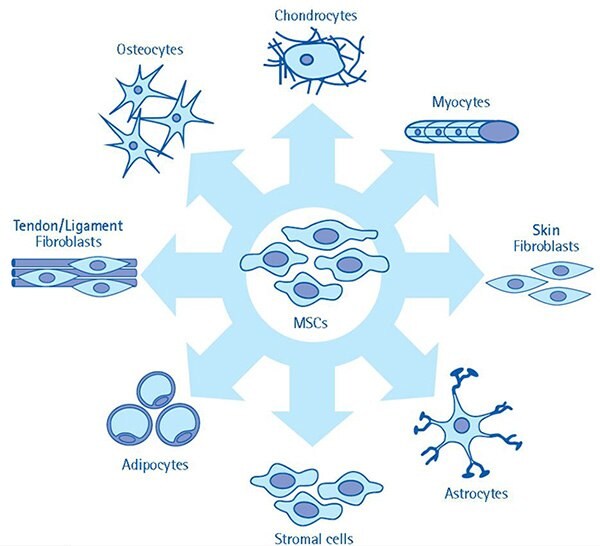
Mesenchymal stem cells differentiate into cells derived from all three lineages, such as:
- Skin, neurons, glial cells derived from ectoderm
- Bone, cartilage, fat and muscle cells derived from mesoderm
- Hepatocytes, pancreatic cells derived from endoderm
What are the methods used to isolate mesenchymal stem cells?
Human mesenchymal stem cells are isolated based on their tissue origin. Majorly, two methods are being used:
- Ficoll density gradient method: Used to isolate mesenchymal stem cells from bone marrow, peripheral blood and synovial fluid.4
- Collagenase digestion method: Used to isolate mesenchymal stem cells from adipose, dental, foreskin, endometrium, Wharton’s Jelly and placenta.5
What are the characteristics of MSC?
International Society of Cellular therapy has listed specific characteristics of mesenchymal stem cells, which are:6
- Adherence to plastic surface
- Expression of cell surface markers, including CD105, CD73 and CD90
- And lack expression of CD45, CD34, CD14 or CD11b, CD79, or CD19 and human leucocyte antigen-DR (HLA-DR)
Which media is used to expand mesenchymal stem cells in culture?
Optimized and high quality MSC expansion media are available including Human Mesenchymal-LS Expansion Medium, Human Mesenchymal-XF Expansion and cGMP compliant Stemline® Mesenchymal Stem Cell Expansion Medium.
In addition PLTMax® Human Platelet Lysate is a growth factor rich human derived media supplement that is a superior alternative to fetal bovine serum (FBS) for human mesenchymal stem cell (MSC) culture.
Which media is suitable to differentiate MSC into osteocytes?
OsteoMAX-XF™ is a specially formulated xeno-free media to readily differentiate human mesenchymal stem cells into osteocytes. OsteoMAX-XF was based on patented, bead-based combinatorial technology, that produces mature osteocytes expressing bone-specific and mineralization markers in as little as 7-10 days.
Are there any clinical treatments available using MSC?
No, still they are in clinical trials. The multi-lineage potential and homing ability of MSC offers therapeutic strategies to treat skeletal and neurodegenerative diseases. Considering the secretion of anti-inflammatory molecules and immunoregulatory effects, MSCs are also promising to treat autoimmune and inflammatory diseases.
What are autologous and allogenic MSC and what are its pros and cons?
Why do mesenchymal stem cells show immunosuppression and low immunogenicity?
Mesenchymal stem cells avoid immune response by:
- Expressing low levels of class I MHC molecules and not expressing class II MHC and co-stimulatory molecules (CD80, CD86 and CD40).
- Inhibiting immunogenic activity of T cells, B cell, dendritic cells and natural killer cells either through cell-cell contacts or soluble factors.7

Figure 1. The multi-lineage differentiation potential of mesenchymal stem cells. MSC cultures have the multi-lineage capacity to differentiate towards a variety of cell types. Given the ability of MSCs to give rise to a number of cell types, these cells are highly attractive models for investigation, especially in regenerative medicine applications.
Materials
References
To continue reading please sign in or create an account.
Don't Have An Account?