Secondary Antibodies, Conjugates, and Kits
Secondary Antibodies
Secondary antibodies are polyclonal or monoclonal antibodies that bind to primary antibodies, or antibody fragments, such as the Fc or Fab regions. They are typically labeled with probes that make them useful for detection, purification, or sorting applications. Our polyclonal secondary antibodies are produced from the serum of host animals such as rabbit, goat, and sheep, whereas monoclonal secondary antibodies are produced from primarily mouse hybridoma clones. Secondary antibodies are used in many applications including ELISA, immunopurification, Western Blotting, IHC, IF, flow cytometry, and more.
The specific utility of a secondary antibody depends upon its conjugated probe(s). Probes are molecules that support various detection technologies. The most common detection systems for conjugated secondary antibodies are colorimetric or fluorescent. Colorimetric assays are typically based on the use of alkaline phosphatase (AP) or horseradish peroxidase (HRP) or its derivatives. The biotin avidin (streptavidin) conjugate binding system is often used to amplify the colorimetric signal for AP or HRP. The most common fluorescent assays utilize Fluorescein (FITC), Rhodamine or its derivative, TRITC, Cyanine (Cy3), or Phycoerythrin (R-PE), CF™ or Atto dyes.
We offer a wide variety of secondary antibodies.
Antibody Specificity
Secondary antibodies have been raised to immunoglobulins of various species to provide reagents for visualizing unconjugated primary antibodies bound to antigens in immunoassays. Antibodies to IgG that are whole molecule specific or Fab specific will usually react with all Ig classes, whereas heavy chain specific and Fc specific antibodies will react only with the indicated Ig class. The researcher will need to determine which specificity is best suited to their work. The assay system and the presence of extraneous protein targets that could bind the antibody and give rise to false positive results or high background will affect the choice of reagents. Table A summarizes the various types of secondary antibodies and their uses.
Conjugates
Alkaline Phosphatase Conjugates
Alkaline phosphatase (AP) is an intestinal enzyme that dephosphorylates alcohols, phenols and amines at alkaline pH. It is a 140 kDa homodimer. The optimal pH range for activity is 9.5-10.5.
Alkaline phosphatase conjugates are widely used in immunoassays such as ELISA,2,3 immunohistochemistry and immunocytochemistry,4 and immunoblotting.5 AP conjugates are useful in tissues where endogenous peroxidase activity may generate high background staining with peroxidase conjugates. They are usually more sensitive than peroxidase conjugates, allowing use of higher dilutions, or detection of lower signals, in ELISA or blotting assays. Endogenous alkaline phosphatase activity in tissue sections may be blocked by adding levamisole (L9756) to the substrate buffer.6 Levamisole inhibits all alkaline phosphatase isoenzymes with the exception of intestinal alkaline phosphatase.
Alkaline phosphatase substrates are available to form either soluble or insoluble products.
Alkaline phosphatase conjugates are not recommended for use with intestinal tissue sections or extracts because of endogenous intestinal alkaline phosphatase activity.

References
Peroxidase Conjugates
Horseradish peroxidase (HRP) is an enzyme that specifically reduces hydrogen peroxide in the presence of a proton-donor. HRP is a 40 kDa glycoprotein. The optimal pH range for activity is 6.0-6.5. HRP exhibits good thermal stability (up to 60 0C) and pH stability (4-10). It is inhibited by azide.
Peroxidase conjugates are used in a variety of immunoassays such as ELISA,4,5 immunohistochemistry and immunocytochemistry,6 and immunoblotting.7 HRP conjugates are useful in tissues such as intestine where endogenous alkaline phosphatase activity may generate high background with alkaline phosphatase conjugates. The selection of available peroxidase substrates is wider than that for alkaline phosphatase, and the colors generated are frequently more intense. Endogenous peroxidase activity may be blocked by treating the tissue with an excess of hydrogen peroxide before addition of the conjugate, or by use of other blocking agents such as phenylhydrazine, azide plus nascent hydrogen peroxide, or periodic acid.8 A wide variety of substrates are available to form either soluble or insoluble products.
HRP conjugates are not recommended for use with samples such as blood cells and kidney tubules that contain high levels of endogenous peroxidase activity.
References
Fluorochrome Conjugates
Fluorophores absorb light at one wavelength, the absorption or excitation wavelength, inducing an excited electronic state. This state is unstable, and the molecule quickly returns to the unexcited, or ground, state by emitting light. Due to energy loss, this light is emitted at a longer wavelength (lower energy), which is termed the emission wavelength. The difference between the excitation and emission wavelengths is unique to each fluorophore, and the intensity of excitation and emission drops quickly as the wavelength varies from the maximum. The Fluorescent Dye Properties Table gives the excitation and emission wavelengths and the fluorescent color for several common fluorophores.
Fluorescent dyes are used to detect molecules in a variety of biological applications.1 Unlike many visible dyes, fluorescent dyes generally may be used under physiological conditions and allow staining of living cells and tissues.2 There is comparatively little overlap between emission wavelengths of many fluorophores, allowing staining with two or more fluorescent probes on one sample.2,3 However, this would require that the excitation wavelengths be very similar and the emission wavelengths be considerably different. Tandem dyes such as Quantum Red™ dye, which is a conjugate of two dyes in which the emission wavelength of one dye matches the excitation wavelength of another, can supply an additional color using the same lamp. Some fluorescent dyes stain cell structures directly, such as acridine orange, DAPI, and Hoechst 33258, which stain nucleic acids. Others, such as fluorescein, rhodamine, and phycoerythrin, are conjugated to antibodies, lectins, nucleotides or other biological probes for localization of specific cell or tissue targets.
Fluorochrome conjugates should be protected from light during storage and use.
References
CF™ Dyes
CF dyes are a series of highly water-soluble fluorescent dyes spanning the visible and near-infrared (near-IR) spectrum for labeling antibodies, proteins, nucleic acids, and other biomolecules. Developed by scientists using new breakthrough chemistries, the brightness, photostability, and color selection of CF dyes rival or exceed the quality of other commercial dyes as a result of rational dye design.
The CF dye product line includes reactive CF dyes, labeling kits, CF-labeled secondary antibodies, and other bioconjugates. This collection further expands our broad range of carefully selected secondary antibodies and conjugates, allowing scientists to achieve greater sensitivity, brighter results, and better photostability in immunoassays.
For more information on CF Dyes or to view the fluorescent dye comparison chart, visit sigma-aldrich.com/cfdyes.
Biotin Conjugates
Biotin (Vitamin H) is a cofactor that binds with high affinity (Ka = 1015) to avidin at a ratio of 4:1. The strength of the binding results in an essentially irreversible interaction. This interaction has been exploited for immunolabeling of antigens in histochemical, blotting, and multiwell assays. Biotinylated antibody probes bind to targets on tissue samples, microtiter plates, or membranes. Avidin, conjugated to enzyme,1 fluorochrome,2 or colloidal metal3 binds to multiple sites on the biotinylated probes. Thus the avidin amplifies the signal, resulting in greater sensitivity than that achieved with an antibody-enzyme or antibody-fluorochrome conjugate alone. Visualization may be accomplished by detection of fluorescence, by the colorimetric or chemiluminescent end product of substrate conversion by the attached enzyme, or by microscopic examination. The avidin-biotin system has been used in immunohistology and immunocytology,4 immunoblotting,5 and ELISA.6 Some tissues, such as liver and kidney, contain endogenous biotin which can lead to high background staining when using the biotin-avidin system.8
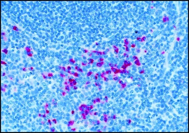
![Goat Anti-Mouse IgM-Biotin Conjugate Frozen section of mouse spleen stained for B cells using Goat Anti-Mouse IgM-Biotin Conjugate (Cat. No. B9265). Visualized using ExtrAvidin ® -Peroxidase (Cat. No. E2886), DAB and nickel chloride, then counterstained with methyl green. 100X shows germinal center formation [From W. Lee, Finch University of Health Science, Chicago Medical School, Department of Microbiology and Immunology, N. Chicago, IL.]](/deepweb/assets/sigmaaldrich/marketing/global/images/technical-documents/articles/protein-biology/elisa/goat-anti-mouse-igm-biotin-conjugate/goat-anti-mouse-igm-biotin-conjugate.jpg)
Figure 2. Frozen section of mouse spleen stained for B cells using Goat Anti-Mouse IgM-Biotin Conjugate (Cat. No. B9265). Visualized using ExtrAvidin® -Peroxidase (Cat. No. E2886), DAB and nickel chloride, then counterstained with methyl green. 100X shows germinal center formation. [From W. Lee, Finch University of Health Science, Chicago Medical School, Department of Microbiology and Immunology, N. Chicago, IL.]
References
Avidin, ExtrAvidin® and Streptavidin Reagents
Avidin has been reported to exhibit non-specific binding to membranes and tissues. For applications where nonspecific binding of avidin is a problem, we offer Streptavidin and ExtrAvidin®.
Avidin, ExtrAvidin® and Streptavidin are available unconjugated or conjugated to enzymes, fluorochromes and colloidal gold for use in immunoassays. Avidin is also available immobilized on agarose for immunoprecipitation or affinity purification procedures.
Avidin
Avidin is a 65 kDa protein found in egg whites. It consists of 4 identical subunits, each with a high-affinity binding site for biotin (Vitamin H). The strength of the binding (Ka = 1015) results in an essentially irreversible interaction. This interaction has been exploited for immunolabeling of antigens in histochemical, blotting, and multiwell assays. Biotinylated probes, which may be secondary antibodies, lectins, or other bioactive compounds, bind to targets on tissue samples, microtiter plates, or membranes. Avidin conjugated to an enzyme, fluorochrome, or other label binds to the biotinylated probes for visualization, either by detection of fluorescence or enzymatic conversion of substrate to produce a visible end product. In this system avidin serves as a secondary probe, attaching to several sites on the primary biotinylated probe and amplifying the signal. Use of an avidin-enzyme conjugate provides further amplification by conversion of substrate by the enzyme, which will continue to produce a visible product until the substrate is exhausted or the reaction is stopped.
ExtrAvidin®
ExtrAvidin® is a modified form of egg white avidin that retains the high affinity and specificity of avidin for biotin, but does not exhibit the nonspecific binding at physiological pH that has been reported for avidin.
Streptavidin
Streptavidin is a form of avidin produced by Streptomyces avidinii that exhibits somewhat less non-specific binding than egg white avidin, although background staining may still sometimes be a problem. Streptavidin is a homotetrameric protein of approximately 60 kDa composed of four identical subunits of approximately 15 kDa each. One molecule of streptavidin binds four molecules of biotin by a non-covalent interaction that is essentially irreversible.
ExtrAvidin® Staining Kits
Available exclusively in our catalog, ExtrAvidin® is a unique form of avidin that combines the high specificity and affinity of avidin for biotin with low non-specific binding at physiological pH. ExtrAvidin® alkaline phosphatase and peroxidase conjugates thus exhibit high sensitivity with low background.
Features and Benefits
References
To continue reading please sign in or create an account.
Don't Have An Account?