Nancy-520 for DNA Detection and Quantitation
Monika Baeumle
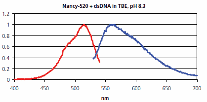
Nancy-520 is a fluorescent stain for double stranded DNA (dsDNA) on agarose electrophoresis gels, with higher sensitivity than ethidium bromide and an easy, fast, and robust staining procedure. It is less mutagenic than Ethidium bromide and SYBR® Green I, according to Ames test. Nancy-520 has an excitation maximum at 520 nm and an emission maximum at 560 nm (Figure 1). Nancy-520 is provided as a 5000× stock solution (500 μL), which is sufficient for 50 agarose gels. The limit of detection is 0.5 ng/band of dsDNA. In addition, Nancy-520 can be used to determine dsDNA concentrations in solution

Figure 1.Normalized fluorescence excitation (red) and emission (blue) spectra of Nancy-520 in the presence of dsDNA, measured in a cuvette on a Varian Cary® Eclipse fluorescence spectrophotometer with 2 mL of TBE, pH 8.3. The excitation spectrum was measured at a fixed emission wavelength of 560 nm, and the emission spectrum was measured at a fixed excitation wavelength of 520 nm.
DNA Staining
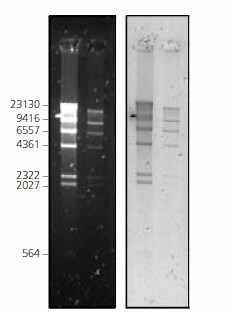
Nancy-520 can be used in a standard post-electrophoresis staining application for DNA on agarose gels. After the electrophoresis run, the gel is stained for 1 hour in the dark. The fluorescence image of the gel can be directly detected after a short rinse (Figure 2). Alternatively, Nancy-520 can also be used for pre-electrophoresis staining by simply adding the dye to the liquid agarose before pouring the gel. The gel can be imaged directly after the electrophoresis run without any further steps. Nancy-520 allows more detection possibilities than SYBR® Green. Detection is performed by illuminating the gel on a UV screen or using a Dark Reader®, and imaging the gel using a CCD-camera (e.g., KODAK® Gel-Logic-100) with a 535 nm or 590 nm band-pass filter, or a Polaroid® camera. Alternatively, a laser-scanner can be used (e.g., Fuji® FLA-3000) with a 473 nm excitation setting and 520 nm emission filter, or with a 532 nm excitation setting and 580 nm emission-filter. Other imaging systems are possible with similar excitation sources and emission filter settings.

Figure 2DNA marker (Lambda DNA Hind III digest) at 2 different concentrations, was separated on a 1% agarose gel, stained with Nancy-520 post-electrophoresis and imaged under 2 different conditions. Left: λex UV-Screen (300 nm)/λem 590 nm bandpass filter/CCD camera. Right: Laser-scanner Fuji FLA-3000/λex 532 nm/λem 580 nm cut-off filter.
DNA Quantitation in Solution
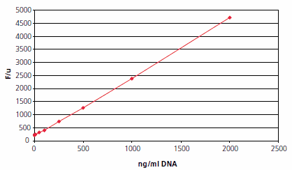
Nancy-520 can be used to determine dsDNA concentrations in solution. This application can be performed in a glass-bottomed 96 well plate, using known concentrations of dsDNA as a standard. Nancy-520 has a linear detection range between 0-2 μg/mL of DNA (Figure 3).

Figure 3.Quantitation of DNA (Lambda DNA Pst I Digest) in solution using Nancy-520,performed in a 96 well plate. Fluorescence was measured on a laser-scanner (FLA-3000,Fuji) with 532 nm excitation and 580 nm emission filter. Different concentrations of DNA in TE buffer, pH 7.5, and their corresponding fluorescence values are shown.
Materials
To continue reading please sign in or create an account.
Don't Have An Account?