Detection of Vancomycin-Induced Subacute Nephrotoxicity Using Kidney Toxicity Multiplex Assays
Nephrotoxicity is often difficult to assess with traditional biomarkers that only increase after substantial kidney damage. Therefore, novel biomarkers are being studied to help detect acute nephrotoxicity. Multiplex kidney toxicity biomarker assays allow researchers to investigate multiple biomarkers of renal damage in a very small sample volume, reducing the need to infer kidney injury based on a single biomarker while minimizing time and costs. Read on to see how MILLIPLEX® multiplex kidney toxicity assays were used to detect vancomycin-induced subacute nephrotoxicity in rat models.
How Do You Assess Kidney Toxicity?
In the pharmaceutical and chemical industries, the kidney is routinely assessed during preclinical safety evaluations. The kidney is an important central detoxification organ because there is an extraordinary exposure of renal tissue to drugs, reactive metabolites, or environmental chemicals. This exposure can lead to cell damage, primarily due to high blood flow, xenobiotic metabolism, or high clearance.1 The frequency at which drug-induced nephrotoxicity occurs, relative to other drug-induced toxicities, is 2% to 20%.2 The reason for this range may be due to the difficulty of assessing kidney toxicity. Kidney toxicity, also known as nephrotoxicity or renal toxicity, is usually assessed by using traditional markers, which are well-known to be insensitive, as is the case with blood urea nitrogen (BUN) and serum creatinine (SCr). Although both are direct measurements of renal function, increases in serum concentrations of these biomarkers occur only after substantial renal injury, generally after the loss of two-thirds of the nephrons’ functional capacity.3 In the case of acute kidney injury (AKI), the degeneration of renal tissue can occur after days or even weeks of exposure.4,5
What Are Kidney Toxicity Biomarkers?
For improved detection of acute nephrotoxicity, a set of novel urinary kidney toxicity biomarkers was approved by the U.S. Food and Drug Administration (FDA), European Medicines Agency (EMA), and Japan’s Pharmaceuticals and Medical Devices Agency (PMDA). The Predictive Safety Testing Consortium (PSTC) - composed of these government entities and leading pharmaceutical companies, together with the nonprofit Critical Path Institute (C-Path) - led the discovery of a large number of transcriptional biomarkers and subsequent evaluation of 23 urinary proteins in the rat.6,7 Some of these markers have been accepted (“qualified”) by the regulators for the detection of AKI in preclinical rodent studies for up to 14 days. Table 1 gives an overview of the most relevant biomarkers measured in this study. Because the majority of the studies performed by the PSTC members included study time points of 1, 3, 7, and 14 days, for an improved risk assessment, it was determined to be absolutely necessary to generate further information about the kinetics of the markers, as well as to test their utility in subacute (28 days) or subchronic (commonly 90 days) preclinical rodent studies. Therefore, this study focused on addressing how these urinary biomarkers perform after 28 days of treatment with a model nephrotoxic compound, vancomycin.8 This compound is a glycopeptide antibiotic that has been previously shown to affect primarily the tubules in cases of vancomycin-induced kidney injury.7,8
Multiplexed Detection of Vancomycin-Induced Subacute Nephrotoxicity
In this study, 120 Wistar rats were divided into groups of 10 animals (5 female, 5 male), and were treated with vancomycin at 2 doses (low dose: 50 mg/kg body weight [bw]; high dose: 300 mg/kg bw) for up to 28 days. The candidate proteins were measured in urine on days 4, 8, 15, and 29 using the MILLIPLEX® Rat Kidney Toxicity Magnetic Bead Multiplex Panels to assess their ability to detect drug-induced renal damage. For interpretation of the data, standard clinical/chemical and histopathological observations were performed. An earlier study with the same set of samples and similar methodology has already been reported.8 The data obtained from the two studies were consistent and reproducible. The ability to predict compound-dependent renal damage and distinguish it from acute renal failure, often associated with other risk factors, has the potential to positively affect drug development productivity.
Materials and Methods
Animal Studies
The 28-day rat study was performed according to Good Laboratory Practice and in compliance with the German Law on the Protection of Animals (German Animal Welfare Act, Article 8a). Before the studies were conducted, a dose range finding study was carried out under the same conditions as planned for the main studies. Significant results of the dose range finding study have been reported.8 Ten-week-old Wistar rats, purchased from Charles River Laboratories (Sulzbach, Germany), were randomly divided into 12 groups of 10 animals (5 females, 5 males). Animals were individually housed in type III isolated ventilated Makrolon® cages with a 12-hour light-dark cycle.
Before treatment, male rats weighed 287 ± 9.0 g and female rats weighed 195 ± 5.8 g. Rats were treated with vancomycin (Ratiopharm, Ulm, Germany) at either a low dose of 50 mg/kg or a high dose of 300 mg/kg. Vancomycin was diluted in water and administered intraperitoneally (i.p.) daily for 7 days and thereafter only once per week. The control animals received an equal volume of 0.9% saline for 7 days by daily i.p. injection, followed by administration twice per week. On day 4, 8, 15, and 29, blood, urine, and organ tissue samples were collected from all individual animals (n=10 for each group; 5 male, 5 female).
The animals were housed in individual metabolism cages for 18 hours (fasted with free access to water). Urine was collected under cooled conditions and stored at -80 °C until the urinary biomarkers were measured. Blood samples were taken by puncture of the sublingual vein under light isoflurane anesthesia. Blood samples and aliquots of urine were immediately used for routine clinical chemistry analyses. Frozen aliquots of urine for the determination of urinary protein biomarkers were all measured in parallel. The animals were sacrificed by CO2 asphyxiation, and organs were collected and fixed in formalin for histopathological examination.
Clinical Pathology
Urine and plasma analyses were carried out on an ADVIA 1650 Autoanalyzer and Clinitek 100 Reflection Spectrophotometer (Siemens Medical Solution Diagnostics GmbH, Bad Nauheim, Germany) using standard protocols for the determination of multiple parameters, based on the recommendations from Weingand and colleagues and according to the manufacturer’s instructions.11,12
Kidney Toxicity Biomarker Analysis
MILLIPLEX® Rat Kidney Toxicity Magnetic Bead Panels using the Luminex® xMAP® platform were used for the safety biomarker analysis. This analysis requires a platform that will reliably quantify proteins from preclinical samples with high sensitivity and specificity. ELISAs are an alternate platform for accurate quantification; however, their low throughput is a major drawback. In addition, preclinical kidney toxicity studies typically use small rodents, resulting in limited available sample volumes.
To overcome these limitations, several emerging technology platforms, such as the Luminex® xMAP® platform and others, have been developed and compared, making it possible to measure several proteins in a single sample robustly and rapidly.10,13 Multiplexed analysis offers significant advantages concerning time, reagent cost, sample requirements, and the amount of data that can be generated. Combined with high-quality antibody reagents and assay development expertise, multiplexed immunoassays on the Luminex® xMAP® platform can deliver the sensitivity, reproducibility, dynamic range (pg/mL to ng/mL), throughput, and robustness demanded for quantitative biomarker analysis.
The MILLIPLEX® Rat Kidney Toxicity Magnetic Bead Panel 1 includes multiplexed assays for Clusterin, GSTα, IP-10, KIM-1, Osteopontin, TIMP-1, and VEGF. This kit may be used for the simultaneous quantification of all or any combination of the analytes in urine and requires only 25 μL of 1:2 diluted urine sample volume.
The MILLIPLEX® Rat Kidney Toxicity Magnetic Bead Panel 2 includes multiplexed assays for AGP, Albumin, β-2-Microglobulin, Cystatin C, EGF, and Lipocalin-2/NGAL. This kit may be used for the simultaneous quantification of all or any combination of the analytes in dilute urine samples and requires only 25 μL of 1:500 diluted urine sample volume.
The results presented below illustrate the application of MILLIPLEX® Rat Kidney Toxicity Magnetic Bead Panels 1 and 2 in a preclinical study.
Statistical Analysis
Urinary multiplexing assay results were calculated from an 8-point standard curve and were normalized against the appropriate urinary creatinine value. Statistical analysis was performed by analysis of variance (ANOVA) and Tukey test. Values significantly different from control are indicated as: *p < 0.05, **p < 0.01, and ***p < 0.001.
Renal Toxicity Analysis Results
Renal histopathology of this cohort of rats was previously reported.8 Macroscopic enlargement of kidneys in high dose-treated (HD) animals was observed. Histopathological observation of tissues prepared from formalin-fixed paraffin-embedded (FFPE) blocks (hematoxylin/eosin stained) showed that vancomycin treatment caused general tubular alterations in HD animals after 28 days of treatment. HD animals showed multifocal, massive to severe tubular degeneration in combination with a moderate to severe tubular dilation.8
KIM-1 Immunohistochemistry
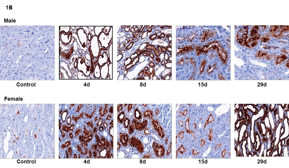
Gender-specific differences in the location of KIM-1 were identified at all time points in the HD groups (Figure 1A). An increase in protein level was observed up to Day 8. Cellular regeneration up to Day 15 led to a reduction in KIM-1. On Day 29, when massive tubular damage was again observed, KIM-1 was also up-regulated (Figure 1B).


Figure 1.(A) Gender-specific differences in the localization of KIM-1 at all time points in the HD animals (Day 8 shown). (B) Time course of KIM-1 expression in male and female rats treated with high dose (300 mg/kg) vancomycin.
Serum Creatinine and Blood Urea Nitrogen
The classic serum markers for detecting the loss of renal function, SCr and BUN, showed strong effects after 7 and 28 days of 300 mg/kg (HD) vancomycin treatment (Figure 2).

Figure 2.Significant increases were observed at several time points in the high dose-treated group of both genders in BUN and SCr. No changes in low-dose-treated animals could be detected. ANOVA + Dunnett p-values: * <0.05, ** <0.01, *** <0.001.
Analysis of Urinary Kidney Biomarkers
Fourteen urinary protein biomarkers were measured using the MILLIPLEX® Rat Kidney Toxicity Magnetic Bead Panel 1 and Panel 2: AGP, Albumin, β2M, Clusterin, Cystatin C, EGF, GSTα, IP-10, KIM-1, NGAL, OPN, TIMP-1, and VEGF.
No urinary markers showed significant differences in the low-dose-treated (LD) groups throughout the 28-day time course. In the HD groups, increases in urinary Clusterin, KIM-1, NGAL, Osteopontin (OPN), and TIMP-1 (Figure 3) reflected the renal regeneration and strength of the vancomycin-induced response, correlating with histopathological findings (data not shown).8

Figure 3.Urinary kidney biomarker analysis by MILLIPLEX® Rat Kidney Toxicity Magnetic Bead Multiplex Panels to predict vancomycin-induced kidney damage: Clusterin, KIM-1, NGAL, Osteopontin (OPN), and TIMP-1. Data represent individual animals and mean values are represented by a line. Statistical significance was determined by ANOVA + Dunnett-Test: *p<0.05, **p<0.01, ***p<0.001.
Interpretation of AGP, Albumin, Cystatin C, and EGF levels was limited by gender differences, illustrating significant and related alterations to vancomycin treatments in the male groups but not in the female groups (Figure 4).

Figure 4.Urinary kidney biomarker analysis by MILLIPLEX® Rat Kidney Toxicity Magnetic Bead Multiplex Panels to predict vancomycin-induced kidney damage: AGP, Albumin, Cystatin C, and EGF. Data represent individual animals and mean values are represented by a line. Statistical significance was determined by ANOVA + Dunnett-Test: *p<0.05, **p<0.01, ***p<0.001.
β2M, GSTα, IP-10, and VEGF demonstrated minimal changes in this study and therefore were not predictive of vancomycin-induced renal damage (Figure 5).

Figure 5.Urinary kidney biomarker analysis by MILLIPLEX® Rat Kidney Toxicity Magnetic Bead Multiplex Panels to predict vancomycin-induced kidney damage: β2M, GST, IP-10, and VEGF. Data represent individual animals and mean values are represented by a line. Statistical significance was determined by ANOVA + Dunnett-Test: *p<0.05, **p<0.01, ***p<0.001.
Conclusion
In this study, acute nephrotoxicity biomarkers were investigated for their ability to detect renal damage in a vancomycin-induced toxicity study in rats over a period of 28 days at 4 time points. MILLIPLEX® Rat Kidney Toxicity Magnetic Bead Panels, based on Luminex® xMAP® multiplexing technology, were employed and compared to histopathology and immunohistochemistry data.
The data demonstrate the high accuracy and predictivity of several of these new markers, even after 28-day subacute treatment with one well-described nephrotoxin, vancomycin, which caused a very distinct kidney tubular damage in rats. The data were consistent with previous studies of subacute vancomycin treatment.8 The classic markers, BUN and SCr, showed significant increases after 7 days of treatment (Day 8 groups, Figure 2). No significant changes were observed on Day 4. In contrast, significant changes in urinary Clusterin, KIM-1, NGAL, Osteopontin (OPN), and TIMP-1 levels could be detected on Day 4, demonstrating a higher sensitivity and specificity of these novel urinary protein biomarkers (Figure 3).
Furthermore, the strong increase of KIM-1 after vancomycin treatment reflected a severe insult on the renal proximal tubular cells and delivered additional information on the location of damage, compared with the traditional parameters alone. Interestingly, a group of urinary markers (AGP, Albumin, Cystatin C, and EGF) showed a gender-specific detection of vancomycin-induced kidney toxicity only in the male groups and not in the female groups (Figure 4). Based on the known toxicity of vancomycin, these biomarker results, predicting primarily tubular damage, are consistent with the literature on kidney injury.7,8
Many questions remain with regards to the utility of these novel nephrotoxicity biomarkers. It is of major interest to discover the optimum timeline of biomarker excretion, especially against the background of the high regenerative properties of renal tissue. It is also of interest to identify any potential recovery from renal injury after cessation of drug exposure, and this can be assessed based on excretion of the urinary biomarkers.
In addition, besides age dependency in biomarker excretion, which has recently been shown, gender differences in urinary biomarker excretion have been illustrated in this study.14 These differences could influence the sensitivity and/or specificity of some of the markers. Therefore, before implementing these assays into preclinical studies, further investigations are required to differentiate between treatment-induced effects and general variations in the biomarker expression pattern across a population. Further investigations should also address excretion and the ability to discriminate gender-specific toxic insults induced by xenobiotics.
The Luminex® xMAP® technology-based MILLIPLEX® Rat Kidney Toxicity Assays can accommodate a broad context of study designs. The complexity of accumulating standardized preclinical data, interpreting the data, and ranking the new markers is a compelling reason to use these biomarker panels, which are comprised of several markers, as opposed to inferring kidney injury based on a single biomarker or a small number of biomarkers. Practically, these panels enable researchers to measure multiple nephrotoxicity biomarkers in a very small sample volume, enhancing the predictive power of preclinical models while minimizing time, animal, and reagent costs.
This research was conducted at EMD Serono’s Department of Non-Clinical Safety and Imperial College in London, UK. Data was provided by Philip Hewitt, Ph.D., Tobias Christian Fuchs, and Esther Johann in 2017.
Related Products
Bead-based multiplex immunoassays enable a precise, multiparametric analysis of the concurrent processes that underlie toxicity. No single biomarker has the power to tell you what you need to know to profile the effect on your model, and our growing MILLIPLEX® portfolio provides protein biomarkers in a multiplex format, using the Luminex® xMAP® bead-based technology.
Our MILLIPLEX® multiplex toxicity biomarker assays make us the leading partner for toxicity testing research.
Materials
For Research Use Only. Not For Use In Diagnostic Procedures.
References
如要继续阅读,请登录或创建帐户。
暂无帐户?