Skip to Content
Merck
CN

DUO80102

Sigma-Aldrich

Duolink® In Situ Brightfield Mounting Medium

Sign Into View Organizational & Contract Pricing

Select a Size


About This Item

UNSPSC Code:
12352200
Technical Service
Need help? Our team of experienced scientists is here for you.
Let Us Assist
Technical Service
Need help? Our team of experienced scientists is here for you.
Let Us Assist

product line

Duolink®

technique(s)

proximity ligation assay: suitable

storage temp.

20-25°C

Application

Experiments conducted using Duolink in situ reagents can detect and visualize protein interactions, protein expression levels and post translational modifications at the single molecule level in fixed cells and tissue samples.

To perform a complete Duolink in situ experiment you will need two primary antibodies (IHC or IF validated) that recognize two target epitopes. Additional reagents required include a pair of PLA probes, one PLUS and one MINUS , your choice of Detection Reagents. Optional reagents include Wash Buffers and Mounting Medium.

Analysis is carried out using standard immunofluorescence assay equipment. HRP/Novared is also available for bright field detection. Quick and reliable quantification can be performed using the Duolink Image Tool.

Application Note
Two primary antibodies raised in different species are needed. Test your primary antibodies (IgG-class, mono- or polyclonal) in a standard immunofluorescence (IF) or immunohistochemistry (IHC) assay to determine the optimal fixation, blocking, and titer conditions.
Duolink in situ reagents are suitable for use on fixed cells, cyt-spin cells, cells grown on slide, formalin-fixed, paraffin embedded (FFPE), or tissue (fresh or frozen). No minimum number of cells is required.

Find answers to commonly asked question on our Duolink FAQ page

Legal Information

Duolink is a registered trademark of Merck KGaA, Darmstadt, Germany

Signal Word

Danger

Hazard Classifications

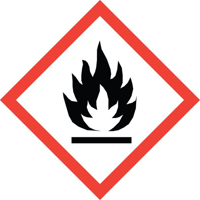
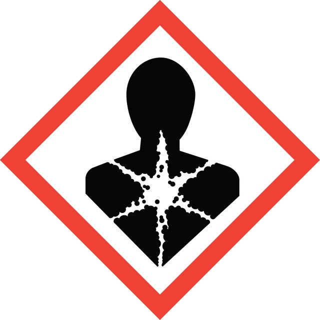
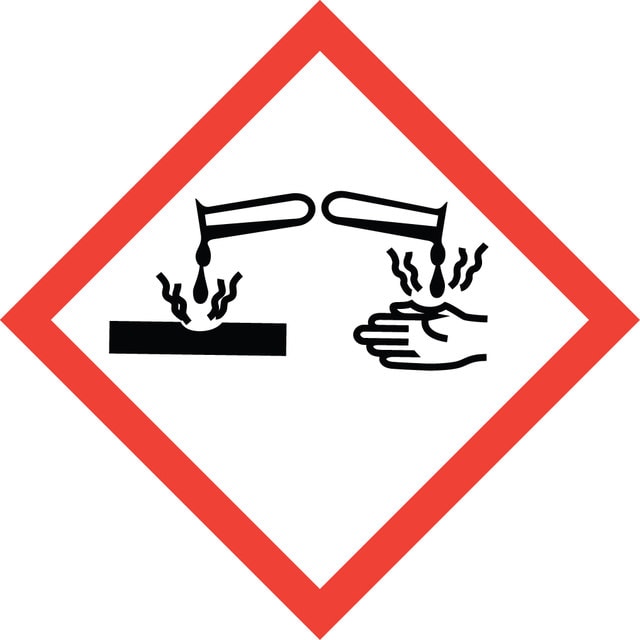
Acute Tox. 4 Dermal - Acute Tox. 4 Inhalation - Aquatic Chronic 3 - Asp. Tox. 1 - Eye Dam. 1 - Flam. Liq. 3 - Skin Irrit. 2 - STOT RE 2 - STOT SE 3

Target Organs

hearing organs, Respiratory system

Storage Class Code

3 - Flammable liquids

WGK

WGK 3

Flash Point(F)

73.4 °F - closed cup

Flash Point(C)

23 °C - closed cup

Regulatory Information

新产品
This item has

Choose from one of the most recent versions:

Certificates of Analysis (COA)

Lot/Batch Number

Don't see the Right Version?

If you require a particular version, you can look up a specific certificate by the Lot or Batch number.

Already Own This Product?

Find documentation for the products that you have recently purchased in the Document Library.

Visit the Document Library

Maria B Bagh et al.
Nature communications, 8, 14612-14612 (2017-03-08)
Defective lysosomal acidification contributes to virtually all lysosomal storage disorders (LSDs) and to common neurodegenerative diseases like Alzheimer's and Parkinson's. Despite its fundamental importance, the mechanism(s) underlying this defect remains unclear. The v-ATPase, a multisubunit protein complex composed of cytosolic
Annabel Christ et al.
Developmental cell, 22(2), 268-278 (2012-02-22)
Sonic hedgehog (SHH) is a regulator of forebrain development that acts through its receptor, patched 1. However, little is known about cellular mechanisms at neurulation, whereby SHH from the prechordal plate governs specification of the rostral diencephalon ventral midline (RDVM)
Lei Zhu et al.
The Prostate, 73(2), 219-226 (2012-07-19)
PSA is the most useful prostate cancer marker. However, its levels are increased also in some non-malignant conditions. In circulation, the majority of PSA is complexed with protease inhibitors, including α(1) -antichymotrypsin (ACT). The proportion of the PSA-ACT complex is
Yoshihiro Akimoto et al.
Clinical proteomics, 8(1), 15-15 (2011-09-22)
The objective of the present study is to identify proteins that change in the extent of the modification with O-linked N-acetylglucosamine (O-GlcNAcylation) in the kidney from diabetic model Goto-Kakizaki (GK) rats, and to discuss the relation between O-GlcNAcylation and the
Agata Zieba et al.
Clinical chemistry, 56(1), 99-110 (2009-11-21)
The in situ proximity ligation assay (PLA) allows a protein or protein complex to be represented as an amplifiable DNA molecule. Recognition is mediated by proximity probes consisting of antibodies coupled with oligonucleotides. Upon dual binding of the proximity probes

Related Content

Applications to detect, quantify and visualize protein-protein interactions, post-translational modifications and low expression protein detection using proximity ligation assay

Our team of scientists has experience in all areas of research including Life Science, Material Science, Chemical Synthesis, Chromatography, Analytical and many others.

Contact Technical Service