Fluorescent Live Cell Imaging of Cytoskeleton Structure Proteins using LentiBrite™ Lentiviral Biosensors
Introduction
Cell analysis of the dynamics of subcellular structures has been revolutionized in the past 15 years by the development and refinement of genetically-encoded fusions between fluorescent proteins (GFP/RFP) and cellular structural proteins. Microtubules are dynamic cytoskeletal filaments composed of tubulin and actin subunits that play central roles during mitosis (in the mitotic spindle) and during interphase as a scaffold for directed kinesin- and dynein-mediated movement of cellular cargo. Several families of microtubule-binding agents, such as taxanes and vinca alkaloids, disrupt microtubule dynamics and cause cell death, and are clinically effective chemotherapeutic agents. Imaging of microtubule dynamics in live cells expressing tubulin tagged with fluorescent proteins has contributed greatly to our understanding of microtubule-binding agents.
We utilized LentiBrite™ lentiviral biosensors to visualize microtubules (α-tubulin), actin microfilaments (β-actin), microfilament cross-link sites (α-actinin), intermediate filaments (vimentin), and focal adhesions (paxillin). We demonstrated that these biosensors, which are validated for use with fixed and live-cell fluorescent microscopy, did not perturb the structures of interest. Localization of the GFP or RFP tagged proteins coincided with the endogenous proteins as determined by immunocytochemistry, and the biosensors displayed characteristic rearrangements upon treatment with known modulators of cytoskeletal structure. Thus, this panel of lentiviral biosensors provides a convenient method for visualization of cytoskeletal structure and dynamics under a variety of physiological and pathological treatment conditions, in both endpoint and real-time live cell imaging modalities.
LentiBrite™ Live Cell Videos
Live Cell Imaging of Autophagy using LentiBrite™ Fluorescent Biosensors
Live Cell Imaging of β-Actin Cytoskeleton Proteins using LentiBrite™ Fluorescent Biosensors
Live Cell Imaging of p62 using LentiBrite™ Fluorescent Biosensors
Methods
Construction of Lentiviral Vectors Encoding Fluorescent Protein Fusions
The cDNAs encoding TagGFP2 and TagRFP were obtained from Evrogen. LentiBrite™ RFP- and GFP-α-tubulin were constructed by cloning the full-length cDNA sequence of the human tubulin α-1B at the C-terminus of the fluorescent protein cDNA. LentiBrite™ RFP- and GFP-β- actin were constructed by cloning the full-length cDNA sequence of human β-actin at the C-terminus of the fluorescent protein cDNA. LentiBrite™ GFP- and RFP vimentin were constructed by cloning the full-length cDNA sequence of the human vimentin at the C-terminus of the fluorescent protein cDNA. LentiBrite™ α-actinin-GFP and -RFP were constructed by cloning the full-length cDNA sequence of human α-actinin-1 isoform b at the N-terminus of the fluorescent protein cDNA. LentiBrite™ paxillin-GFP and -RFP were constructed by cloning the full-length cDNA sequence of human paxillin at the N-terminus of the fluorescent protein cDNA. Constructs were transferred to pCDH-EF1-MCS, a lentiviral vector containing the constitutive, moderately expressing EF1α promoter. 3rd generation HIV-based VSV-G pseudotyped lentiviral particles were generated using the pPACKH1 Lentivector Packaging System.
Cell Seeding and Lentiviral Transduction
Cells in growth medium were seeded onto 8-well glass chamber slides for fixed and live cell imaging. Seeding densities were selected to provide for 50-70% confluency after overnight culture (e.g., 20,000-40,000 cells/cm2). The next day after seeding, medium was replaced with fresh growth medium. High-titer lentiviral stock was diluted 1:40 with growth medium, and appropriate volumes of lentivirus were added to the seeded cells to achieve the desired multiplicity of infection (MOI). MOI refers to the ratio of the number of infectious lentiviral particles to the number of cells being infected. Typical MOI values ranged from 10 to 40. Infected cells were then incubated at 37 °C, 5% CO2 for 24 h. 24 h after lentiviral transduction, lentivirus-containing medium was removed and replaced with fresh growth medium. Cells were cultured for another 24-48 h, with media changed every 24 hours. For inhibitor experiments, cells were incubated in the inhibitor of interest at the concentration and for the time indicated in the legend.
Live Cell Imaging and Antibody Staining
For live cell visualization, the growth medium was replaced with Dulbecco’s Modified Eagle Medium (DMEM) containing 10% fetal bovine serum (FBS) and 25 mM HEPES. The chambered cover glass was placed in a temperature-controlled microscope stage insert upon a Leica DMI6000B inverted wide-field fluorescent microscope with a 63X oil-immersion objective lens and illumination/filters appropriate for GFP or RFP visualization. Imaging was initiated as rapidly as possible following the addition of modulators.
For end-point imaging, cells in chamber slides were fixed for 30 minutes at room temperature with 3.7% formaldehyde in Dulbecco’s phosphate-buffered saline (DPBS). During fixation and for all subsequent steps, cells were protected from light to minimize photobleaching. Samples were then rinsed twice with fluorescent staining buffer (DPBS with 2% blocking serum and 0.25% Triton® X-100). Primary antibody in fluorescent staining buffer was added to each well and incubated 1h at room temperature. Samples were then rinsed three times with fluorescent staining buffer before incubation for 1h at room temperature with fluorescent secondary antibody and DAPI (1 μg/mL) in staining buffer. Finally, samples were rinsed twice each with fluorescent staining buffer and DPBS, and slides were coverslipped with mounting medium containing anti-fade reagent and cover glasses.
Results
Improved Biosensor Expression Efficiency with Lentiviral Transduction
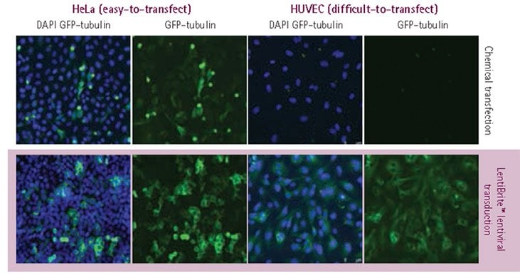
We constructed a panel of lentiviral particles containing genetically encoded fluorescent markers for cytoskeletal elements. By using these lentiviral biosensors, both transfection efficiency and homogeneity were greatly improved relative to chemical transfection with plasmid DNA (Figure 1).

Figure 1.Plasmid vs. lentivirus transfection in easy- and difficult-to-transfect cell types. HeLa cells and HUVECs were transfected with a TagGFP2-tubulin-encoding construct, either utilizing plasmid DNA in conjunction with a lipid-based chemical transfection reagent, or using LentiBrite™ lentiviral particles. Images were obtained via wide-field fluorescent imaging with a 20X objective lens (blue = DAPI nuclear counterstain, green = GFP-tubulin). Lentiviral transduction resulted in higher transfection efficiency (particularly for HUVEC, for which plasmid transfection was unsuccessful) and GFP-tubulin signal of more uniform fluorescence intensity.
Panel of lentiviruses encoding live cell biosensors for cytoskeletal elements

Figure 2.LentiBrite™ lentiviral biosensors localize to specific cytoskeletal elements. (A) GFP-paxillin highlights focal adhesions in REF-52 cells. (B) α-actinin-RFP in REF-52 cells displays a striated filamentous pattern corresponding to actin microfilament cross-link sites. (C) β-actin-RFP in REF-52 cells displays a similar filamentous pattern to that of α-actinin, but with a pattern of continuous intensity. (D) RFP-α-tubulin in HUVECs displays a pattern of microtubule filaments radiating from the microtubule organizing center (white arrow). (E) GFP-vimentin in human MSCs highlights the intermediate filaments.
Specificity and Localization of Lentivirally Delivered Biosensors
As genetically encoded biosensors can be prone to improper localization, due either to a propensity of the particular fluorescent protein to aggregate or to the overexpressed biosensor over-saturating the structure of interest, we sought to confirm the specificity of lentivirally delivered biosensor localization. We found that lentiviral delivery of fluorescent protein-tagged cytoskeletal markers enabled accurate detection of the appropriate cytoskeletal elements, as determined by 1) redistribution of the biosensor upon treatment of the cells with specific inhibitors of the cytoskeletal structure, 2) similar distribution of the biosensor to endogenous protein as determined by immunofluorescence, and 3) live cell imaging of cytoskeletal dynamics.

Figure 3.Localization of RFP-tagged tubulin following modulator treatment. HUVECs were lentivirally transduced with RFP-tubulin biosensors at an MOI of 40. Transduced cells were subjected to the following treatments for 4 h: (A) untreated, (B) treated with 1 μM paclitaxel, or (C) treated with 25 μM nocodazole. Cells were fixed, mounted, and imaged by wide-field fluorescence microscopy. Cells treated with paclitaxel displayed bundled microtubules that curved around the nucleus, whereas cells treated with nocodazole contained no evident microtubule structures.

Figure 4.Fluorescent protein expression and co-localization with fluorescent antibody staining. HeLa cells were transduced with TagRFP-vimentin biosensors at an MOI of 20, and 72 h later, either left untreated or treated with 25 μM nocodazole. Cells were subsequently fixed, immunostained with an anti-vimentin antibody, and imaged by wide-field microscopy. Untreated cells displayed a filamentous vimentin network, in contrast to a clumped perinuclear distribution in cells treated with nocodazole. In both cases, the fluorescently tagged protein (top and middle rows) colocalized with staining obtained with anti-vimentin antibody (bottom row).

Figure 5.Live cell time-lapse imaging of β-actin lentivirally-transduced cells. REF-52 cells were lentivirally transduced with TagRFP-β-actin biosensors, and imaged by oil-immersion widefield microscopy in real time. The cells were dosed with 2 μM cytochalasin D, and imaging was immediately initiated, with images obtained every 30 s for 30 min. Still-frame captures demonstrated transition of fine filamentous organization to a granular pattern in the vicinity of the nucleus, and diffuse distribution at the cellular periphery.
Conclusion
Lentiviral biosensors enable convenient transduction of easy- and difficult-to-transfect cell types with fluorescent protein-tagged subcellular markers. These LentiBrite™ pre-packaged lentiviral particles enable higher efficiency and more homogeneous expression of introduced proteins compared to non-viral transfection methods. We have demonstrated the use of GFP- or RFP-tagged cytoskeletal markers in both fixed and live cell microscopy applications. The encoded proteins displayed co-localization with antibody staining and appropriate redistribution upon treatment with known modulators of cytoskeletal structure. LentiBrite™ biosensors provide a ready-to-use solution for researchers seeking to fluorescently visualize the presence, absence or trafficking of a protein, under normal, abnormal, diseased, or induced cellular states.
Materials
To continue reading please sign in or create an account.
Don't Have An Account?