Merck
CN
HomeCell Counting & Health AnalysisMTT Assay Protocol for Cell Viability and Proliferation

MTT Assay Protocol for Cell Viability and Proliferation

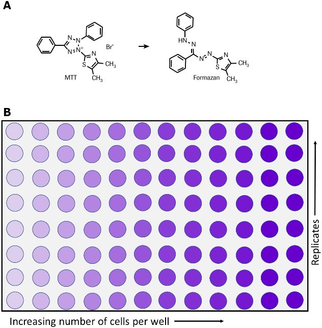
The MTT assay is used to measure cellular metabolic activity as an indicator of cell viability, proliferation and cytotoxicity. This colorimetric assay is based on the reduction of a yellow tetrazolium salt (3-(4,5-dimethylthiazol-2-yl)-2,5-diphenyltetrazolium bromide or MTT) to purple formazan crystals by metabolically active cells (Figure 1).6,7,35 The viable cells contain NAD(P)H-dependent oxidoreductase enzymes which reduce the MTT to formazan.36 The insoluble formazan crystals are dissolved using a solubilization solution and the resulting colored solution is quantified by measuring absorbance at 500-600 nanometers using a multi-well spectrophotometer. The darker the solution, the greater the number of viable, metabolically active cells.

This non-radioactive, colorimetric assay system using MTT was first described by Mosmann, T et al.1 and improved in subsequent years by several other investigators.2-6 The Cell Proliferation Kit I (MTT) is an optimized MTT assay kit containing ready to use reagents, does not need washing steps or additional reagents. It is a quantitative assay that allows rapid and convenient handling of a high number of samples. The Cell Proliferation Kit I (MTT) can be used for multiple applications, such as,

  • Quantification of cell growth and viability.1,3,5-7
  • Measurement of cell proliferation in response to growth factors, cytokines and nutrients.1-3,6,8-12 (see figure 3).
  • Measurement of cytotoxicity. Examples are the quantification of tumor necrosis factor-a or -b effects 13,14 (see figure 2) or macrophage induced cell death15,16 and the assessment of cytotoxic 17-34 or growth inhibiting agents such as inhibitory antibodies.
  • To study cell activation.4
Metabolism of MTT to a formazan salt and 96-well plate

Figure 1.Metabolism of MTT to a formazan salt by viable cells as shown in a chemical reaction (A) and in a 96-well plate (B).

Cell Proliferation Kit I (MTT) Components (Product No. 11465007001)

MTT Reagent

  • Ready to use, non-sterile
  • 5 vials containing 5 ml MTT (3-[4,5-dimethylthiazol-2-yl]-2,5-diphenyltetrazolium bromide) (1X), at 5 mg/mL in phosphate-buffered saline (PBS, 806552)

Solubilization Solution

  • 1X, ready-to-use
  • 3 bottles with 90 mL

Assay Protocol to Measure Cytotoxicity

Additional Reagents Required:

  • Culture medium, e.g., RPMI 1640 (R0883) containing 10% heat inactivated FCS (fetal calf serum, 12106C), 2 mM glutamine (G6392) and 1 μg/mL actinomycin C1 (actinomycin D, A9415).
  • If an antibiotic is to be used, additionally supplement media with penicillin/streptomycin or gentamicin
  • Tumor necrosis factor-α, human (hTNF-α) (10 μg/mL), sterile (T6674).

Protocol:

For the determination of the cytotoxic effect of human tumor necrosis factor-α (hTNF-α, T6674) on WEHI-164 cells (mouse fibrosarcoma, 87022501) (see Figure 2).

  1. Preincubate WEHI-164 cells at a concentration of 1 × 10⁶ cells/ml in culture medium with 1 μg/mL actinomycin C1 for 3 h at 37 °C and 5-6.5% CO2.
  2. Seed cells at a concentration of 5 × 10⁴ cells/ well in 100 μl culture medium containing 1 μg/ml actinomycin C1 and various amounts of hTNF-α (final concentration e.g., 0.001–0.5 ng/mL) into microplates (tissue culture grade, 96 wells, flat bottom).
  3. Incubate cell cultures for 24 h at +37 °C and 5-6.5% CO2.
  4. After the incubation period, add 10 μl of the MTT labeling reagent (final concentration 0.5 mg/ml) to each well.
  5. Incubate the microplate for 4 h in a humidified atmosphere (e.g., +37 °C, 5-6.5% CO2).
  6. Add 100 μl of the Solubilization solution into each well.
  7. Allow the plate to stand overnight in the incubator in a humidified atmosphere (e.g., +37 °C, 5-6.5% CO2).
  8. Check for complete solubilization of the purple formazan crystals and measure the absorbance of the samples using a microplate (ELISA) reader. The wavelength to measure absorbance of the formazan product is between 550 and 600 nm according to the filters available for the ELISA reader, used. The reference wavelength should be more than 650 nm.
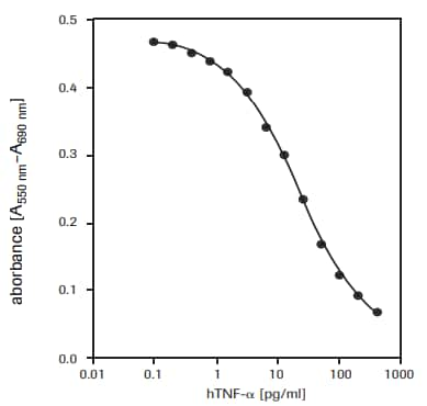
Determination of the cytotoxic activity

Figure 2.Determination of the cytotoxic activity of recombinant human TNF-α (hTNF-α) on WEHI-164 cells (mouse fibrosarcoma) using MTT assay.

Assay Protocol to Measure Cell Growth

Additional Reagents Required

  • Culture medium (e.g., DMEM, Product No. D5671) containing 10% heat inactivated FCS (fetal calf serum, Product No. 12106C)
  • 2 mM glutamine (Product No. G6392)
  • 0.55 mM L-arginine (Product No. A8094
  • 0.24 mM L-asparagine-monohydrate (Product No. A4284)
  • 50 μM 2-mercaptoethanol (Product No. M3148), 
  • HT-media supplement (Product No. H0137) (1×), containing 0.1 mM hypoxanthine and 16 μM thymidine. If an antibiotic is to be used, additionally supplement media with penicillin/streptomycin or gentamicin. 
  • Interleukin-6, human (hIL-6, Product No. SRP3096) (200,000 U/mL, 2 μg/mL) sterile

Protocol

For the determination of human interleukin-6 (hIL-6) activity on 7TD1 cells (mouse-mouse hybridoma, DSMZ, ACC 23) (see fig. 3).

  1. Seed 7TD1 cells at a concentration of 2 × 10³ cells/well in 100 μL culture medium containing various amounts of IL-6 [final concentration e.g., 0.1-10 U/mL (0.001-0.1 ng/mL)] into microplates (tissue culture grade, 96 wells, flat bottom).
  2. Incubate cell cultures for 4 days at +37 °C and 5-6.5% CO2.
  3. After the incubation period, add 10 μL of the MTT labeling reagent (final concentration 0.5 mg/mL) to each well.
  4. Incubate the microplate for 4 h in a humidified atmosphere (e.g., +37 °C, 5-6.5% CO2).
  5. Add 100 μL of the Solubilization solution into each well.
  6. Allow the plate to stand overnight in the incubator in a humidified atmosphere (e.g., +37 °C, 5-6.5% CO2).
  7. Check for complete solubilization of the purple formazan crystals and measure the spectrophotometrical absorbance of the samples using a microplate (ELISA) reader. The wavelength to measure absorbance of the formazan product is between 550 and 600 nm according to the filters available for the ELISA reader, used. The reference wavelength should be more than 650 nm.
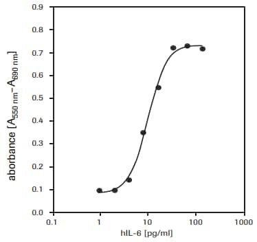
Proliferation of 7TD1 cells

Figure 3.Proliferation of 7TD1 cells (mouse-mouse hybridoma) in response to recombinant human interleukin-6 (hIL-6) using MTT assay.

Similar Assays

XTT assay and WST-1 assay can also be used for measuring cell viability and proliferation.

Products
Loading

References

1.
Mosmann T. 1983. Rapid colorimetric assay for cellular growth and survival: Application to proliferation and cytotoxicity assays. Journal of Immunological Methods. 65(1-2):55-63. https://doi.org/10.1016/0022-1759(83)90303-4
2.
Tada H, Shiho O, Kuroshima K, Koyama M, Tsukamoto K. 1986. An improved colorimetric assay for interleukin 2. Journal of Immunological Methods. 93(2):157-165. https://doi.org/10.1016/0022-1759(86)90183-3
3.
Denizot F, Lang R. 1986. Rapid colorimetric assay for cell growth and survival. Journal of Immunological Methods. 89(2):271-277. https://doi.org/10.1016/0022-1759(86)90368-6
4.
Gerlier D, Thomasset N. 1986. Use of MTT colorimetric assay to measure cell activation. Journal of Immunological Methods. 94(1-2):57-63. https://doi.org/10.1016/0022-1759(86)90215-2
5.
Hansen MB, Nielsen SE, Berg K. 1989. Re-examination and further development of a precise and rapid dye method for measuring cell growth/cell kill. Journal of Immunological Methods. 119(2):203-210. https://doi.org/10.1016/0022-1759(89)90397-9
6.
Vistica VT, Skehan P, Scudiero D, Monks A, Pittman A, Boyd MR. 1991. Tetrazolium-based assays for cellular viability: a critical examination of selected parameters affecting formazan production. Cancer Res. 51(10):2515-20.
7.
Maehara Y, Anai H, Tamada R, Sugimachi K. 1987. The ATP assay is more sensitive than the succinate dehydrogenase inhibition test for predicting cell viability. European Journal of Cancer and Clinical Oncology. 23(3):273-276. https://doi.org/10.1016/0277-5379(87)90070-8
8.
Heeg K, Reimann J, Kabelitz D, Hardt C, Wagner H. 1985. A rapid colorimetric assay for the determination of IL-2-producing helper T cell frequencies. Journal of Immunological Methods. 77(2):237-246. https://doi.org/10.1016/0022-1759(85)90036-5
9.
Hooton, Gibbs J, Paetkan U C. 1985. Interaction of interleukin 2 with cells: quantitative analysis of effects.. J Immunol. 1352464–2473.
10.
Mosmann T, Fong T. 1989. Specific assays for cytokine production by T cells. Journal of Immunological Methods. 116(2):151-158. https://doi.org/10.1016/0022-1759(89)90198-1
11.
Ohno M, Abe T. 1991. Rapid colorimetric assay for the quantification of leukemia inhibitory factor (LIF) and interleukin-6 (IL-6). Journal of Immunological Methods. 145(1-2):199-203. https://doi.org/10.1016/0022-1759(91)90327-c
12.
Berg K, Hansen MB, Nielsen SE. 1988. J. Interferon Res.. 8((suppl. 1)):S. 67.
13.
Green LM, Reade JL, Ware CF. 1984. Rapid colormetric assay for cell viability: Application to the quantitation of cytotoxic and growth inhibitory lymphokines. Journal of Immunological Methods. 70(2):257-268. https://doi.org/10.1016/0022-1759(84)90190-x
14.
Espevik T, Nissen-Meyer J. 1986. A highly sensitive cell line, WEHI 164 clone 13, for measuring cytotoxic factor/tumor necrosis factor from human monocytes. Journal of Immunological Methods. 95(1):99-105. https://doi.org/10.1016/0022-1759(86)90322-4
15.
Ferrari M, Fornasiero MC, Isetta AM. 1990. MTT colorimetric assay for testing macrophage cytotoxic activity in vitro. Journal of Immunological Methods. 131(2):165-172. https://doi.org/10.1016/0022-1759(90)90187-z
16.
van de Loosdrecht AA, Nennie E, Ossenkoppele GJ, Beelen RH, Langenhuijsen MM. 1991. Cell mediated cytotoxicity against U 937 cells by human monocytes and macrophages in a modified colorimetric MTT assay. Journal of Immunological Methods. 141(1):15-22. https://doi.org/10.1016/0022-1759(91)90205-t
17.
Cole S. 1986. Rapid chemosensitivity testing of human lung tumor cells using the MTT assay. Cancer Chemother. Pharmacol.. 17(3): https://doi.org/10.1007/bf00256695
18.
Carmichael J, DeGraff WG, Gazdar AF, Minna JD, Mitchell JB. 1987. Evaluation of a tetrazolium-based semiautomated colorimetric assay: assessment of chemosensitivity testing. Cancer Res. 47(4):936-42.
19.
Twentyman P, Luscombe M. 1987. A study of some variables in a tetrazolium dye (MTT) based assay for cell growth and chemosensitivity. Br J Cancer. 56(3):279-285. https://doi.org/10.1038/bjc.1987.190
20.
Park JG, Park BS, Steinberg SM, Carmichael J, Collins JM, Minna JD, Gazdar AF. 1987. Chemosensitivity testing of human colorectal carcinoma cell lines using a tetrazolium-based colorimetric assay. Cancer Res. 475875-9.

21.
1988. FFPS News. Oryx. 22(4):246-250. https://doi.org/10.1017/s0030605300022481
22.
Pieters R, Huismans D, Leyva A, Veerman A. 1988. Adaptation of the rapid automated tetrazolium dye based (MTT) assay for chemosensitivity testing in childhood leukemia. Cancer Letters. 41(3):323-332. https://doi.org/10.1016/0304-3835(88)90294-7
23.
McHaLe A, McHaLe L. 1988. Use of a tetrazolium based colorimetric assay in assessing photoradiation therapy in vitro. Cancer Letters. 41(3):315-321. https://doi.org/10.1016/0304-3835(88)90293-5
24.
Edmondson JM, Armstrong LS, Martinez AO. 1988. A rapid and simple MTT-based spectrophotometric assay for determining drug sensitivity in monolayer cultures. Journal of Tissue Culture Methods. 11(1):15-17. https://doi.org/10.1007/bf01404408
25.
Faircloth GT, Stewart D, Clement JJ. 1988. A simple screening procedure for the quantitative measurement of cytotoxicity to resting primary lymphocyte cultures. Journal of Tissue Culture Methods. 11(4):201-205. https://doi.org/10.1007/bf01407314
26.
Campling BG, Pym J, Galbraith PR, Cole SP. 1988. Use of the mtt assay for rapid determination of chemosensitivity of human leukemic blast cells. Leukemia Research. 12(10):823-831. https://doi.org/10.1016/0145-2126(88)90036-7
27.
Pieters R, Huismans D, Leyva A, Veerman A. 1989. Comparison of the rapid automated MTT-assay with a dye exclusion assay for chemosensitivity testing in childhood leukaemia. Br J Cancer. 59(2):217-220. https://doi.org/10.1038/bjc.1989.44
28.
Twentyman PR, Fox NE, Rees JKH. 1989. Chemosensitivity testing of fresh leukaemia cells using the MTT colorimetric assay. Br J Haematol. 71(1):19-24. https://doi.org/10.1111/j.1365-2141.1989.tb06268.x
29.
Plumb JA, Milroy R, Kaye SB. 1989. Effects of the pH dependence of 3-(4,5-dimethylthiazol-2-yl)-2,5-diphenyl-tetrazolium bromide-formazan absorption on chemosensitivity determined by a novel tetrazolium-based assay. Cancer Res. 494435–4440.
30.
Alley MC, Scudiero DA, Monks A, Hursey ML, Czerwinski MJ, Fine DL, Abbott BJ, Mayo JG, Shoemaker RH, Boyd MR. 1988. Feasibility of drug screening with panels of human tumor cell lines using a microculture tetrazolium assay. Cancer Res. 48589–601.
31.
Heo DS, Park JG, Hata K, Day R, Herberman RB, Whiteside TL. 1990. Evaluation of tetrazolium-based semiautomatic colorimetric assay for measurement of human antitumor cytotoxicity. Cancer Res. 503681–3690.
32.
Ruben RL, Neubauer RH. 1987. Semiautomated colorimetric assay for in vitro screening of anticancer compounds. Cancer Treat Rep. 711141–1149..
33.
Rubinstein LV, Shoemaker RH, Paull KD, Simon RM, Tosini S, Skehan P, Scudiero DA, Monks A, Boyd MR. 1990. Comparison of In Vitro Anticancer-Drug-Screening Data Generated With a Tetrazolium Assay Versus a Protein Assay Against a Diverse Panel of Human Tumor Cell Lines. JNCI Journal of the National Cancer Institute. 82(13):1113-1117. https://doi.org/10.1093/jnci/82.13.1113
34.
1991. Announcement. Utilitas. 3(2):330-330. https://doi.org/10.1017/s0953820800001254
35.
Slater T, Sawyer B, Sträuli U. 1963. Studies on succinate-tetrazolium reductase systems. Biochimica et Biophysica Acta. 77383-393. https://doi.org/10.1016/0006-3002(63)90513-4
36.
Berridge M, Tan A. 1993. Characterization of the Cellular Reduction of 3-(4,5-dimethylthiazol-2-yl)-2,5-diphenyltetrazolium bromide (MTT): Subcellular Localization, Substrate Dependence, and Involvement of Mitochondrial Electron Transport in MTT Reduction. Archives of Biochemistry and Biophysics. 303(2):474-482. https://doi.org/10.1006/abbi.1993.1311
Sign In To Continue

To continue reading please sign in or create an account.

Don't Have An Account?