High-Performance Thin-Layer Chromatography: A Fast and Efficient Fingerprint Analysis Method for Medicinal Plants
Ginsenosides are triterpene saponins. Most ginsenosides are composed of a dammarane skeleton (17 carbons in a four-ring structure) with various sugar moieties (e.g. glucose, rhamnose, xylose and arabinose) attached to the C- 3 and C-20 positions.
Over 30 ginsenosides have been identified and classified into two categories:
• 20(S)- protopanaxadiol (PPD) (Rb1, Rb2, Rb3, Rc, Rd, Rg3, Rh2, Rs1)
• 20(S)-protopanaxatriol (PPT) (Re, Rf, Rg1, Rg2, Rh1). The difference between PPTs
and PPDs is the presence of a carboxyl group at the C-6 position in PPDs.

Figure 1.
Moreover, several rare ginsenosides, such as the ocotillol saponin F11 (24-R-pseudoginsenoside) and the pentacyclic oleanane saponin Ro (3,28-o-bisdesmoside) have also been identified. Asian ginseng (Panax ginseng) is commonly known as the true ginseng.
In this article, HPTLC methods suitable for the analysis of ginsenosides are presented, using CAMAG equipment, MilliporeSigma TLC plates, analytical standards and extract reference materials. The extract reference materials are manufactured by HWI Analytik and exclusively distributed by MilliporeSigma.
Detection of ginsenosides in the HPTLC fingerprint of different Panax species (roots and root extracts) is obtained by following the HPTLC methods of the Ph. Eur. monograph,1 USP DSC 2015 monograph2 and the method of the HPTLC Association (International Association for the Advancement of HPTLC)3 by comparison of the RF values and colors of reference substances and matching zones in the root extract. Depending on the regulation followed, one of the three methods of identification can be used.
Recommended CAMAG Devices:
Automatic TLC Sampler (ATS 4), Automatic Developing Chamber (ADC 2), TLC Visualizer 2, Chromatogram Immersion Device 3, TLC Plate Heater 3, and visionCATS
Derivatization Reagent:
Anisaldehyde1 or sulfuric acid reagent2,3
Sample:
0.015 g/mL extract (HWI extract) in 70% methanol1 Note: Deviation from methods 2 and 3 for the sample preparation
Standards:
Standard solutions of ginsenosides were prepared in a concentration of 0.5 mg/mL in methanol.
Note: Deviation from method 3 for the application volumes
Chromatography Following USP <203> 4:
Stationary phase: HPTLC Si 60 F254 20 x 10 cm (MilliporeSigma)
Sample application: 4 µL each of test solution and 2 µL of standards are applied as 8 mm bands, 8 mm from lower edge, 20 mm from the left edge1, 3
Note: Deviation from method 3 for the sample preparation and the application volume of reference and test solutions
4 µL each of test solution and 4 µL of standards are applied as 8 mm bands, 8 mm from lower edge, 20 mm from the left edge2
Note: Deviation from method 2 for the sample preparation
Developing solvent: Ethyl acetate – water – butanol 25:50:100 (v/v/v) - upper layer1
Dichloromethane – ethanol – water 70:45:6.5 (v/v/v)2
Chloroform – ethyl acetate – methanol – water 15:40:22:9 (v/v/v/v)3
Development: In the ADC 2 with an unsaturated chamber and after conditioning at 33% relative humidity for 10 min using a saturated solution of magnesium chloride1
Development is performed with ADC 2, saturated for 20 minutes with the developing solvent (filter paper). Prior to the development the plate is conditioned for 10 min to a relative humidity of 33% (with a saturated solution of MgCl2).2, 3
Developing distance: 70 mm (from lower edge)1, 2; 80 mm (from lower edge)3
Plate drying: 5 min in a stream of cold air
Derivatization: The plate is immersed (immersion speed: 3 cm/s, immersion time: 0 s) into anisaldehyde reagent (mixture of 0.5 mL of p-anisaldehyde, 10 mL of glacial acetic acid, 85 mL of methanol, and 5 mL of sulfuric acid) with the Chromatogram Immersion Device 3 and heated for 5 min at 105°C.1
The plate is immersed (immersion speed: 3 cm/s, immersion time: 0 s) into sulfuric acid reagent (10% in methanol) with the Chromatogram Immersion Device 3 and heated for 5 min at 100 °C.2, 3
Evaluation: Documentation under method 3 is with 100 °C white light1, 2, 3 and UV 366 nm2, 3 after derivatization with the TLC Visualizer 2
Results and Discussion:
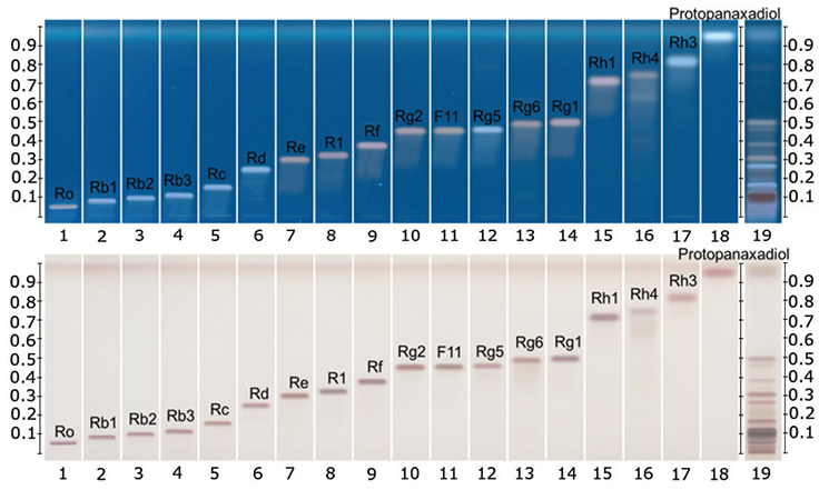
HPTLC chromatograms of ginsenoside standards and a Panax ginseng root extract
HPTLC chromatograms after derivatization. Tracks
1-17: ginsenosides, track 18: protopanaxadiol, track 19: Panax ginseng root extract (article no.: 05115001 batch: HWI01294)

Method 1:According to the Ph. Eur.
HPTLC chromatograms under white light after derivatization

Method 2:According to the Tienshi ginseng method from
HPTLC chromatograms under UV 366 nm and under white light after derivatization

Method 3:According to the HPTLC Association
HPTLC chromatograms under UV 366 nm and under white light after derivatization.
HPTLC chromatograms of plants containing ginsenosides (different Panax species)
P. ginseng, P. quinquefolium, P. notoginseng, P. japonicus, P. vietnamensis roots and root extracts were collected and analyzed. The P. ginseng root extract (article no.: 0511-50-01 batch: HWI01294) was used as botanical reference material to identify Asian ginseng (the ginsenoside Rf should be present and F11 absent).

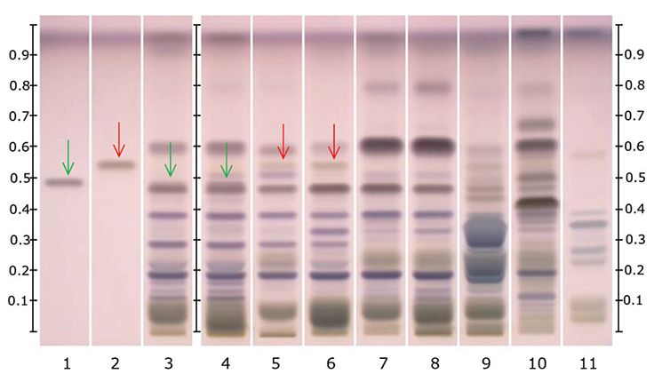
Method 1: According to the Ph. Eur.
HPTLC chromatogram under white light after derivatization.
Track 1: ginsenoside Rf (green arrow); 2: ginsenoside F11 (red arrow); 3: P. ginseng root extract (article no.: 0511-50-01 batch: HWI01294, ginsenoside Rf highlighted with the green arrow); 4: P. ginseng root (ginsenoside Rf highlighted with the green arrow); 5: P. quinquefolium root extract (American ginseng, ginsenoside F11 highlighted with the red arrow);
6: P quinquefolium root (American ginseng, ginsenoside F11 highlighted with the red arrow); 7: P. notoginseng root extract;
8: P. notoginseng root; 9: P. japonicas root; 10: P. vietnamensis root; 11: wild Vietnamese ginseng root.

Method 2:According to the Tienshi ginseng method from
HPTLC chromatograms under UV 366 nm and under white light after derivatization

Method 3:According to the HPTLC Association
HPTLC chromatograms under UV 366 nm and under white light after derivatization.
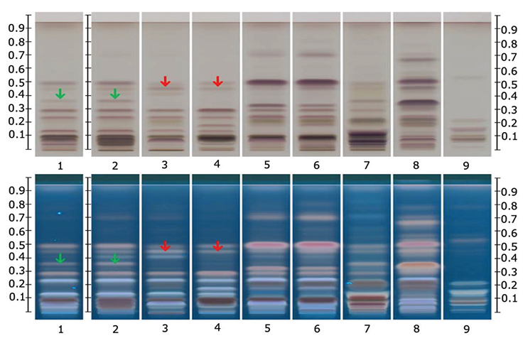
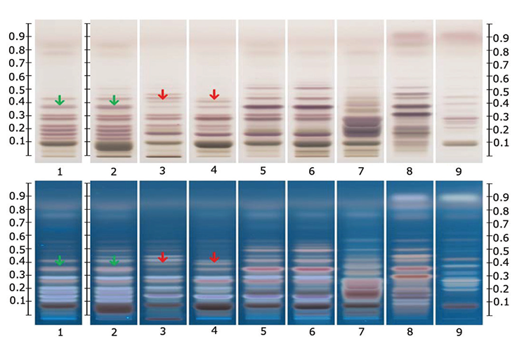
Method 2 & 3 Parameter:
HPTLC chromatograms under white light and UV 366 nm after derivatization with ginsenoside Rf (green arrows) and ginsenoside F11 (red arrows).
Track 1: P. ginseng root extract (article no.: 05115001 batch: HWI01294, ginsenoside Rf highlighted with the green arrows); 2: P. ginseng root (ginsenoside Rf highlighted with the green arrows); 3: P. quinquefolium root extract (American ginseng, ginsenoside F11 highlighted with the red arrows); 4: P quinquefolium root (American ginseng, ginsenoside F11 highlighted with the red arrows); 5: P. notoginseng root extract; 6: P. notoginseng root; 7: P. japonicas root; 8: P. vietnamensis root;
9: wild Vietnamese ginseng root.
All shown methodologies are suitable for detection of ginsenosides in different Panax species. Ginsenoside Rf is unique to Asian ginseng while F11 is found exclusively in American ginseng. Thus the Rf/F11 ratio is used as a phytochemical marker to distinguish American ginseng from Asian ginseng. In the botanical reference material used (Panax ginseng root extract, article no.: 0511-50-01) the presence of Rf and absence of F11 could be confirmed with all methods and it is therefore suited for identification of Asian ginseng.
Methods 1 and 2 have the advantage in that they are in accordance with official monographs in the pharmacopoeias (Ph. Eur. resp. USP). Method 3 is an alternative method provided by the HPTLC Association. The method is improved for the separation of Rf and F11 to better distinguish American and Asian ginseng. The derivatization with sulfuric acid reagent (methods 2 and 3) leads to different colored zones under UV 366 nm, useful for identification.
Materials
References
To continue reading please sign in or create an account.
Don't Have An Account?