Merck
CN
HomeCell-Based AssaysNeurite Outgrowth Assays

Neurite Outgrowth Assays

In Vitro Neurite Outgrowth Assays
AXIS™ Axon Isolation Devices
Neural Cell Lines and Culture Media
Neuroscience Antibodies
References

Introduction

Neurite Outgrowth is a process wherein developing neurons produce new projections as they grow in response to guidance cues. Nerve growth factors, or neurotrophins, are one family of such stimuli that regulate neurite growth1. Dynamic neurite outgrowth during development results in the formation of a complex neuronal architecture that results in the establishment of the functional nervous system and brain2. Understanding the biology of neurite outgrowth can shed light on mechanisms underlying certain neurodegenerative diseases3. In order to investigate neurite outgrowth, researchers need an effective methods for growing neurons in culture, stimulating the cells with neurogenic compounds, such as growth factors, and monitoring the growing neurites.

In Vitro Neurite Outgrowth Assays

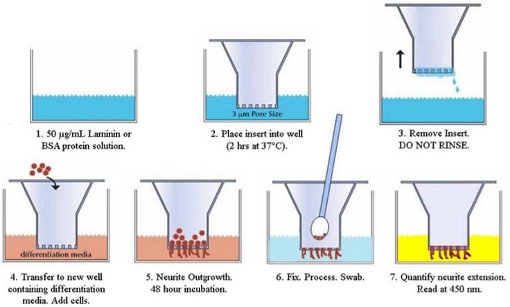
Neurite Outgrowth Assays have been optimized and developed for characterization and quantification of neurite formation, composition, and behavior in response to chemical agents and growth conditions. The assay technique uses Millicell® semi-porous culture inserts (1uM, 3uM) and optimized reagents to easily analyze neurite outgrowth using a common colorimetric or chemiluminescence detection.
Loading
Neurite Outgrowth Assay Protocol

Figure 1. Neurite Outgrowth Assay Protocol. The assay is a simple, efficient, and versatile system for the quantitative determination of compounds that influence neurite formation and repulsion. The microporous filter allows for biochemical separation and purification of neurites and cell bodies for detailed molecular analysis of protein expression, signal transduction processes and identification of drug targets that regulate neurite outgrowth or retraction processes. With this system, it is possible to screen numerous biological and pharmacological agents simultaneously, directly evaluate adhesion and guidance receptor functions responsible for neurite extension and repulsion, as well as analyze gene function in transfected cells.

AXIS™ Axon Isolation Devices

AXIS™ Axon Isolation Devices are slide-mounted microfluidic devices that analyzes neurite outgrowth by spatially controlling addition of growth factors, toxins, and other reagents. Neurite outgrowth is restricted to narrow, parallel channels, and the resultant outgrowth or collapse behavior is easily observed under a microscope. The result is a powerful platform for the study of somas, neurites or synaptic formation.

Key Highlights

  • Organize, visualize, and characterize neuronal cell culture.
  • Detect protein expression with better spatial resolution.
  • Isolate cell bodies from axons through fluidics.
  • Reduce time and expense through optimized protocols and QC validated products.
  • Superior performance over in-house protocols.
  • Optically clear transparent, inert, non-toxic, and nonflammable polymer mold.
  • Available in 150 μm, 450 μm, 900 μm, or 6-well.

Illustration of the Plasma-bonded AXIS® Device

AX50005PB

Illustration of the Plasma-bonded AXIS® Device

Figure 2.Illustration of the Plasma-bonded AXIS™ Device

Effect of the Three Neurotoxins on Axon Growth Visualized by Fluorescence Microscopy

Neurotoxicity Analysis using AXIS™ Axon Isolation Devices

Figure 3.Neurotoxicity Analysis using AXIS™ Axon Isolation Devices. Axons were exposed to 300 μM MPP+, 5 mM acrylamide, and 500 nM rotenone starting at day 4 post plating, exposing the axons to toxins for 3 days. The top row shows axonal extensions that were exposed to toxin, while the bottom row shows the corresponding control axons. The dotted yellow line indicates the end of the microgrooves.

Neural Cell Lines and Culture Media

Primary Neurons

Neural Culture Media

Immortalized Neural Cell Lines

Neurotrophic Factors

Neural Stem Cell Lines and Media

Bioactive Small Molecules

Neuroscience Antibodies

Understanding neurite outgrowth can improve therapeutics for nervous system developmental disorders and neurodegenerative disease. Our extensively validated antibodies for studying neurite outgrowth can help determine the mechanisms of axon growth.

Common Neural Markers

NeuN

b-Tubulin

MAP2

Nestin

Synaptophysin

Pan-Neuronal

Doublecortin

vGlut

Tyrosine Hydroxylase

Explore over 80,000 Antibodies

Neuron

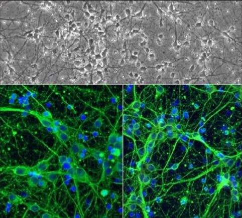
Figure 4.Neuron

References

1.
Davies AM. 2000. Neurotrophins: Neurotrophic modulation of neurite growth. Current Biology. 10(5):R198-R200. https://doi.org/10.1016/s0960-9822(00)00351-1
2.
Tosney KW, Landmesser LT. 1985. Development of the major pathways for neurite outgrowth in the chick hindlimb. Developmental Biology. 109(1):193-214. https://doi.org/10.1016/0012-1606(85)90360-4
3.
Millecamps S, Julien J. 2013. Axonal transport deficits and neurodegenerative diseases. Nat Rev Neurosci. 14(3):161-176. https://doi.org/10.1038/nrn3380
Sign In To Continue

To continue reading please sign in or create an account.

Don't Have An Account?