Blood Brain Barrier Antibody Penetration Assay Using Millicell® Microwell Plates
What is the Blood Brain Barrier?
The blood brain barrier (BBB) is a protective barrier that separates the central nervous system from compounds in circulating blood. It allows or blocks certain substances in the blood, such as antibodies, from entering the human brain. Because it prevents molecules from entering the brain, designing drugs, antibodies, and immunotherapies that target cells in the brain and can pass through the BBB is critical.
Section Overview

Figure 1. Schematic representation of the blood brain barrier.
Antibody therapy has become an important novel tool in neuroscience, neuro-oncology, and immunotherapies. These immunotherapies work through targeting the surface antigens that are expressed on cancer tumors and triggering the body’s immune response1. However, the BBB system is difficult to mimic in vitro and is often studied using animal models.
Here we demonstrate a scalable and high throughput assay using BBB assembloids and Millicell® Microwell 96-well plates in vitro. Our multicellular BBB models are highly reproducible assembloids comprised of brain endothelial cells, astrocytes, and pericytes and rely on self-aggregation. This method allows direct cell-to-cell interactions, closely modeling the in vivo BBB properties.

Figure 2.Representation of BBB models in vitro.
Millicell® Microwell Plates Attributes
Millicell® Microwell 96-well plates are a ready-to-use tool that supports high throughout and reliable 3D culture. The plates do not require an external solid extracellular matrix (ECM), as the microwells in the plate contain a polyethylene glycol (PEG) hydrogel, which is water based and promotes less diffraction during imaging. The PEG hydrogel allows for ease of cell seeding and assay development. Millicell® Microwell plates also contain a media exchange port away from the microwells so users can easily exchange media without microtissue loss.
Spheroids or organoids seeded in the microwells form in a single focal plane and can be scaled up for the easy implementation of automated high throughput imaging and screening applications. Because the BBB assembloids were generated in Millicell® Microwell 96-well plates, there was a 55-fold increase in organoid yield, with more than 5000 datapoints in a single 96-well plate when compared to more traditional suspension culture methods. The assembloids also closely represent the in vivo architecture of the BBB, and thus represent an ideal model for high throughput antibody penetration assays.
How to Choose a Millicell® Microwell 96-well Plate
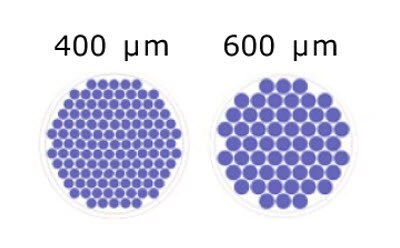
There are two standard designs for different microwell sizes within the wells of Millicell® Microwell 96-well plates. The addition of the microwells allows for scaled up organoid generation depending on your application:

Figure 3.Schematic representation of the microwells/well for each microwell size.
Blood Brain Barrier Antibody Penetration Protocol
- Seed endothelial, pericytes, and astrocytes cell lines simultaneously into the 600m microwells Millicell® Microwell 96-well plate.
- Add culture media through the media exchange port.
- Allow assembloids to self-assemble, compact, grow, and fully develop within 48 hours.
- Image from the bottom of the Millicell® Microwell 96-well plates to monitor assembloid size, roundness, and viability.
- Incubate assembloid with the antibodies of interest; to monitor antibody penetration into the assembloids. For this assay, antibodies that were assessed for penetration were incubated for up to 6 hours.
- Fix and immunnostain on the Millicell® Microwell plate. For this protocol, DAPI (nucleus), P-gP (endothelial cells), NG2 (pericytes), and GFAP (astrocytes) were used, and cell death was monitored with Calcein-AM and Eth heterodimer.
- Evaluate antibody penetration via indirect immunofluorescence with a confocal microscope.
Blood Brain Barrier Antibody Penetration Results
Using the Millicell® Microwell 96-well plates, the BBB assembloids self-assembled into distinct layers. This was visualized through the presence of specific markers for each cell type that were detected directly on the Millicell® Microwell plates. In the BBB assembloids, the endothelial cells (P-gP) surround the pericytes (NG2), which surround a core of astrocytes (GFAP) (Figure 5).

Figure 4.BBB organoid imaging on the Millicell® Microwell plate. A. Brightfield imaging of one Millicell® Microwell plate well with 55 BBB assembloids. B. BBB organoid live/dead staining. Scale bar 600μm.

Figure 5.Immunofluorescence of BBB organoid layers. DAPI (nucleus), P-gP (endothelial cells), NG2 (pericytes), and GFAP (astrocytes). Scale bar 50 μm.
Immunostaining could be directly quantified for each cell line in the layers through imaging performed directly on Millicell® Microwell 96-well plates. The antibodies penetrated the multicellular organoid layers and this novel organoid system allowed for direct cell-cell interactions, similar to what occurs in the BBB in vivo.

Figure 6.Immunofluorescence and quantification of antibody penetration of BBB assembloids. A. untreated control; B. Non-targeting human IgG, endothelial cells were labelled with anti-P-gP; C. Human brain shuttle. Scale bar 100 μm; D) Quantification of antibody penetration through fluorescent intensity.
References
To continue reading please sign in or create an account.
Don't Have An Account?