Array CGH Analysis of Challenging Samples
Read more about
Introduction
In recent years, array-based Comparative Genomic Hybridization (aCGH) has been refined to determine chromosomal changes at progressively higher resolutions1. This evolving technology is, however, hampered by the large DNA input requirement—a minimum of 150,000 copies of a human genome, or 0.5 μg, are generally needed per sample to rocess one CGH array. Often the available tissue collected from a patient, such as small needle biopsies, can limit the scope of downstream analysis. GenomePlex® whole genome amplification (WGA) provides a means to decrease the amount of required DNA for aCGH, in addition to other analyses. The WGA product has also been demonstrated to work on BAC arrays2, 3 and other oligo array platforms4. DNA amplification can expand the number and breadth of applications, such as microarray profiling, to analysis of nanogram quantities of DNA, or even single cells. Furthermore, and perhaps more interesting, GenomePlex WGA enables researchers to representatively amplify the genome from samples that have been fragmented to an average size of less than 1 kb.
Results
100 ng to 1 ng of input DNA

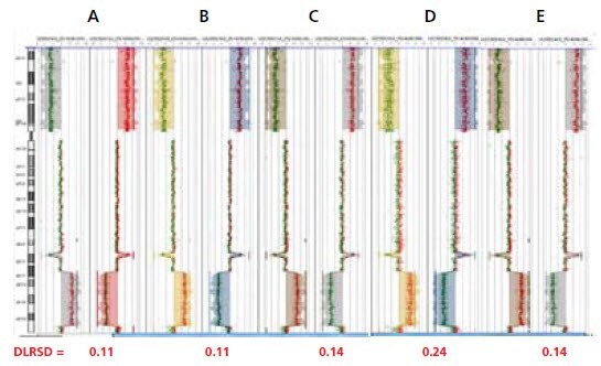
Figure 1.Input titration data from Agilent Human Genome CGH 105A microarrays
Dye swap plots are presented side-by-side in each panel (A-F). Polarity +1 (Cy5-HT29/Cy3-Female) is shown on the left and polarity -1 (Cy5-Female/Cy3-HT29) is shown on the right. These CGH plots compare normal female DNA vs. HT29 human colon carcinoma cell line DNA which has a known deletion across the p arm (horizontal shift to left of zero line), an amplification of the q arm in the q23.3-24.33 region (horizontal shift to right of zero line), and a focal deletion in q23.1. Dye swaps (switching the dye labels between samples) were performed on these samples to demonstrate that the results were not an effect of dye bias. Derivative Log2Ratio Standard Deviation (DLRSD) values are shown beneath plots. Array quality metrics are provided in Table 1.
A: Amplified from 100 ng input
B: Amplified from 50 ng input
C: Amplified from 10 ng input
D: Amplified from 1 ng input
E: Unamplified
Gel image, sonication time course of genomic DNA
Figure 2. 1.2% Agarose gel electrophoresis analysis of fragmented DNAs
DNA fragment size was determined via gel electrophoresis prior to amplification with GenomePlex WGA. Both human female genomic DNA and HT29 genomic DNA yield a high molecular weight intact band (> 12,000 bp), whereas the DNA was variably sonicated for 5 to 120 sec to yield DNA smears of varying sizes. The 90 sec sonication samples (200–1500 bp, lanes 5 and 9) were used in subsequent aCGH analysis.
Lane 1 & 7: Molecular Weight Marker
Lane 2: Intact Female DNA (> 12,000 bp)
Lane 3: Female DNA sonicated 5 sec (400 – 10,000 bp)
Lane 4: Female DNA sonicated 30 sec (250 – 1,500 bp)
Lane 5: Female DNA sonicated 90 sec (200 – 1,200 bp)
Lane 6: Female DNA sonicated 120 sec (150 – 600 bp)
Lane 8: Intact HT29 DNA (> 12,000 bp)
Lane 9: HT29 DNA sonicated 90 sec
GenomePlex WGA can amplify DNA below 1kb with results equivalent to intact DNA.

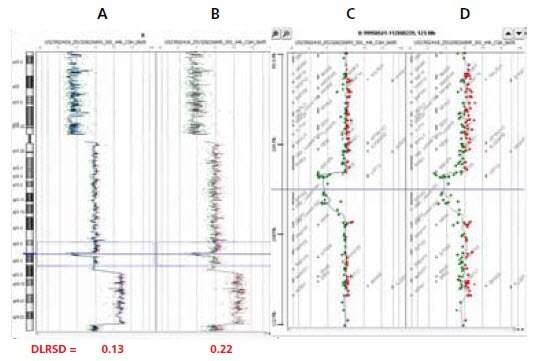
Figure 3.CGH Analytics views of Agilent Human Genome CGH 44B microarrays of chromosome 8 in the human colon carcinoma cell line HT29 vs. normal female
WGA product generated from intact HT29 DNA (A) and sonicated (90sec) HT29 DNA (B) revealed identical known deletion across the 8p arm (horizontal shift to left of zero line), amplification along the 8q arm in the 8q23.3-24.23 region (horizontal shift to right of zero line), and a focal deletion in 8q23.1 (horizontal shift to left of zero line, outlined by dotted blue box). Corresponding zoomed-in gene views for data from both intact DNA (C) and fragmented DNA (D) focusing on Chr8 q22.2-23.1, show a distinct ~1.5 MB deletion (marked as thick line) involving the zinc finger protein ZFPM2 and tumor suppressor LRP12 that have been previously reported in BAC array-based analyses.
A: Chromosome 8 view of amplified intact DNA comparing HT29/Female (50 ng input)
B: Chromosome 8 view of amplified sonicated 90sec DNA comparing HT29/Female (50 ng input)
C: Zoomed-in gene view of panel A (12 MB zoom window)
D: Zoomed-in gene view of panel B (12 MB zoom window)
Identification of a focal deletion with Agilent Human Genome CGH 244K microarrays using either GenomePlex amplified or unamplified DNA.

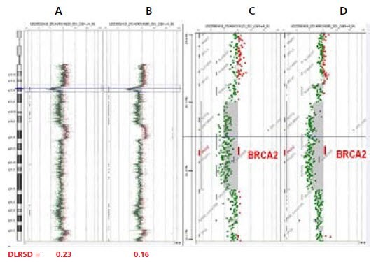
Figure 4.CGH Analytics views of Agilent 244K analyses (5) of a human frozen breast tumor vs. normal female
Scatter plot (chromosome view) produced from 244K microarray analysis reveals a focal deletion in chromosome 13 p13.1 region (horizontal shift to left of zero line, outlined by dotted blue box) in both the amplified (A) and the unamplified (B) samples. Corresponding zoomed-in gene view (C =amplified, D=unamplified) which focuses on a 3.7 MB window within 13p13.1 containing the focal deletions. Multiple deletion patterns involving breast cancer gene BRCA2 (marked with red bar) within the 1.5MB focal deletion across the 13p 13.1 arm were identified by both WGA product and unamplified genomic DNA generated from breast tumor DNA.
A: Breast tumor vs. normal female Chr13, WGA Amplification (50 ng input)
B: Breast tumor vs. normal female Chr13, Unamplified (500 ng input)
C: Zoomed-in gene view of panel A (3.7 MB window)
D: Zoomed-in gene view of panel B (3.7 MB window)
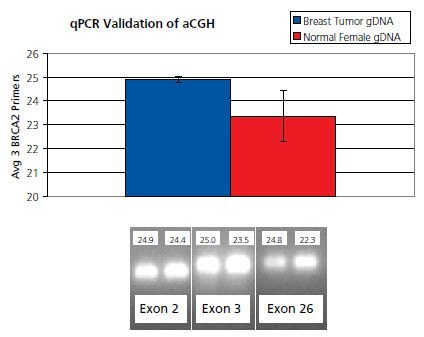
Validation of aCGH breast cancer data using BRCA2 gene-specific quantitative PCR

Figure 5: The average of three SYBR Green quantitative PCR primer sets targeting exons 2, 3 and 26 (6) of the BRCA2 gene demonstrated a 1.54 cycle difference between breast tumor DNA and normal female DNA. This 2.9 fold (2^1.54) drop in representation of the BRCA2 gene from the tumor sample compared to the normal sample validates the 13 p13.1 focal heterozygous deletion.
Conclusions
This amplification method allows for lesser amounts (nanogram quantities) of starting material, and is capable of immortalizing precious samples through successive reamplifications. Aside from foregoing the initial restriction enzyme digestion, WGA DNA can be directly incorporated into the oligo-aCGH workflow without any major deviation from Agilent’s recommended protocol. Amplified material produces high-quality array CGH data that match results from unamplified genomic DNA samples. Future experiments will focus on producing high quality genomic profiles from tissue samples, including FFPE and LCM samples 7.
Materials
References
To continue reading please sign in or create an account.
Don't Have An Account?