NanoFabTx™ Nanoparticle Formulation
Read more about
- The Nanofabtx™ Drug Delivery Formulation Platform
- Nanofabtx™ Polymeric Nanoparticle and Lyposome Synthesis Using the Nanoassemblr®
- Results
- General Protocol for Nanofabtx™ Polymeric Nanoparticle Synthesis Using the Nanoassemblr®
- General Protocol for Nanofabtx™ Mrna Liposome Preparation Using the Nanoassemblr®

Nanoparticle drug delivery formulations overcome many limitations associated with free therapeutics. However, determining the correct formulation can be difficult and often requires lengthy trial-and-error optimization. The NanoFabTx™ drug delivery formulation platform addresses these challenges by providing a highly flexible platform to synthesize, screen, and select the microparticle, nanoparticle, or liposome formulation that best fits your needs. The NanoFabTx™ drug delivery formulations kits and lipid mixes have been developed and tested by our R&D formulation scientists for both small molecule and nucleic acid delivery and include detailed protocols with step-by-step instructions to make synthesis simple.
NANOFABTX™ POLYMERIC NANOPARTICLE AND LYPOSOME SYNTHESIS USING THE NANOASSEMBLR®
The NanoFabTx™ platform has been designed to be synthesis-method agnostic. Nanoparticles can be generated via traditional nanoprecipitation using standard laboratory glassware, or via microfluidics using our NanoFabTx™ microfluidic device kits. Here, we demonstrate our NanoFabTx™ drug formulation screening kits and lipid mixes can also be used with Precision Nanosystems Inc.’s (PNI) NanoAssemblr® and NanoAssmeblr® Ignite microfluidic platform. Various PEG-PLGA nanoparticles were synthesized, optimized, screened, and selected using the NanoFabTX™ PEG-PLGA drug formulation screening kit (917796). Additionally, liposomes containing mRNA were prepared using our NanoFabTx™ DOTAP lipid mix (926027).
RESULTS
NanoFabTx™ PEG-PLGA Nanoparticle Synthesis Using the NanoAssemblr®
The NanoFabTx™ PEG-PLGA drug formulation screening kit (917796) is a ready-to-use nanoformulation kit for the synthesis of PEGylated PLGA nanoparticles. This kit provides four properly selected PEGylated PLGAs and stabilizer, allowing rapid screening of optimal formulation materials for enhanced drug delivery.
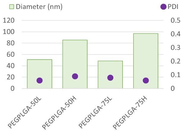
NanoFabTx™ PEG-PLGA FormulationPolymeric nanoparticles synthesized using the NanoFabTx PEG-PLGA drug formulation screening kit and NanoAssemblr®
Nanoparticles of uniform size (PDI ~0.06-0.09) and various formulations were generated using the NanoFabTx™ PEG-PLGA drug formulation screening kit (917796) and PNI’s NanoAssemblr®. The NanoFabTx™ PEG-PLGA drug formulation screening kit consists of four different PEG-PLGA formulations that can be screened and selected to best suit the researcher's needs. Here, nanoparticles of varying formulations were manufactured rapidly and with precise size control, eliminating lengthy trial-and-error optimization. The same flow rate ratio (1:1) and polymer concentration (2.5 wt%) were used to reach formulation.
Precise and Controlled Synthesis OF NanoFabTx™ PEG-PLGA Nanoparticles
Nanoparticle size was controlled by varying manufacturing parameters within the Precision Nanosystems software. A range of nanoparticle sizes (48 nm to 60 nm) were achieved with increased polymer concentration. While polymer concentration directly contributed to nanoparticle size, the PDI remained low, suggesting uniform size distribution.
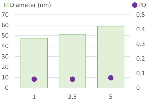
Polymer Concentration (wt%)Controlled synthesis of PEG-PLGA nanoparticles using the NanoFabTx PEG-PLGA drug formulation screening kit and NanoAssemblr®
NanoFabTx™ PEG-PLGA Nanoparticle Size can be Controlled by Altering the Flow Rate Ratio (FRR)
Flow rate ratio (FRR) is another manufacturing parameter that can be used to control nanoparticle size. Here, the flow rate ratio in the PNI software was changed from 1:1 to 1:3 and 1:5, respectively, while maintaining a total flow rate of 8 ml/min. PEG-PLGA nanoparticle size decreased with increased FRR. Nanoparticles generated at each FRR were uniform in size and resulted in a low PDI.
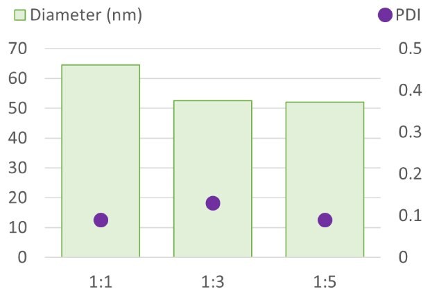
Flow Rate Ratio (Polymer:Stabilizer)NanoFabTx™ PEG-PLGA nanoparticle size can be controlled by flow rate ratio (FRR)
Taking NanoFabTx™ Formulation from Early Discovery to Preclinical Development Using the NanoAssemblr®
The NanoFabTx™ drug delivery formulation platform is designed to aid researchers in their formulation journey—from screening to preclinical development. Here, the NanoFabTx™ PEG-PLGA drug formulation screening kit was used with PNI’s screening and preclinical development NanoAssemblr® devices: the NanoAssemblr® and the NanoAssemblr® Ignite™.
PNI’s NanoAssemblr® is ideal for screening formulations because it requires minimal sample consumption, is easy to use, and rapidly produces nanoparticles of uniform size. The microfluidic technology can also be scaled up across the NanoAssemblr® Platform to accelerate future development. The NanoAssemblr® Ignite platform takes production from initial discovery stages to early preclinical formulation development. Here, NanoFabTx™ PEG-PLGA nanoparticle production by the NanoAssemblr® and NanoAssemblr® Ignite™ devices were compared.

Polymer ConcentrationNanoFabTx™ PEG-PLGA nanoparticles were synthesized using the NanoAssemblr® and NanoAssemblr® Ignite™
PEG-PLGA nanoparticles were generated at various polymer concentrations using the NanoAssemblr® and NanoAssemblr® Ignite™ instruments. Nanoparticle sizes ranged from 50nm to 175nm depending on polymer concentration. The flow rate parameters established using the NanoAssemblr® device were applied to the NanoAssemblr® Ignite™. The results suggest nanoparticles of similar size were generated between the two instruments and that the NanoFabTx™ formulation screening kits can easily be scaled up from early-stage screening to preclinical development.
The results above are indicative of the capabilities and flexibility of the NanoFabTX™ platform. Uniform nanoparticles were generated using the NanoAssemblr® instrument, and nanoparticle size could be adjusted in a controlled and precise manner. Additionally, NanoFabTx™ nanoparticle manufacturing could be scaled up across the NanoAssemblr® Platform.
NanoFabTx™ mRNA Containing DOTAP Liposomes Prepared With the NanoAssemblr®
The NanoFabTx™ DOTAP lipid mix (926027) is designed to prepare specifically sized cationic liposomes for gene delivery applications. The lipid mix contains rationally selected lipids to achieve a specific size range and positive charge for efficient complexation with negatively charged nucleic acids. Here, mRNA containing DOTAP liposomes were prepared using the NanoAssemblr® and NanoFabTx™ DOTAP lipid mix (926027).
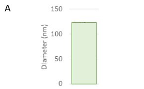

Luciferase reporter mRNA was encapsulated in DOTAP liposomes using the NanoAssemblr® following the protocol provided below. Reproducible mRNA containing DOTAP liposomes were prepared of desired size (approximately 120nm) with an encapsulation efficiency of approximately 90%. Here, an N/P ratio of 5:1 and a flow rate ratio (FRR) of 3:1 was chosen and can be optimized depending on mRNA/nucleic acid and desired liposome size.

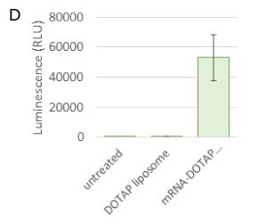
Luciferase Reporter mRNA-Containing Catatonic DOTAP Liposomes: mRNA containing NanoFabTx™ DOTAP liposomes prepared with the NanoAssemblr® Ignite
The in vitro transfection of mRNA-containing DOTAP liposomes was assessed. mRNA-containing DOTAP liposomes were delivered to HeLa cells. After 24 hours, the mRNA containing liposomes and empty liposomes were cytocompatible, resulting in 100% cell viability. Transfection of mRNA was evaluated via luciferase (luminescence) expression. mRNA delivered in the DOTAP liposomes expressed the targeted protein, whereas the untreated cells, and those treated with empty liposomes did not.
The protocol listed below provides step-by-step instructions on mRNA containing DOTAP liposomes using the NanoAssemblr® and NanoFabTX™ DOTAP lipid mix (926027). This protocol was optimized for a specific reporter mRNA strand as a model. It is suggested as a guide for your own optimization, and additional modifications may be necessary for other therapeutics/nucleic acids of interest.
We offer additional NanoFabTx™ formulation screening kits, lipid mixes, and microfluidic device kits. Our R&D scientists have carefully selected and curated each formulation kit and lipid mix to ensure reproducible and unform nanoparticle production to best suit researchers’ drug delivery applications. Each kit has been designed to be preparation method agnostic— nanoparticles can be generated with standard laboratory glassware or NanoAssemblr® instruments following the general protocols described here.
GENERAL PROTOCOL FOR NANOFABTX™ POLYMERIC NANOPARTICLE SYNTHESIS USING THE NANOASSEMBLR®
For material requirements, notes, reagent preparation, and drug quantification, please refer to the protocol included with the NanoFabTx™ PEG-PLGA drug formulation screening kit (917796).
Protocol
- Prime the system
- Turn “on’’ on the Microfluidic system, connect the computer to the microfluidic system, and open the PNI software.
- Insert the micromixing microfluidics cartridge into the microfluidic system.
- Fill one syringe with 1 ml of acetonitrile and fill a second syringe with 1 ml of deionized water.
- Uplift the cartridge holder.
- Load the acetonitrile syringe to one side of the cartridge insert and the water syringe to the other side of the cartridge insert.
- Put down the cartridge holder to its original position.
- Load two 15 ml tubes (labeled waste) to the sample tube holder socket.
- In the PNI software add the following parameters:
o Total Volume: 2 ml
o Flow Rate Ratio (FRR): 1:1
o Total Flow Rate (TFR): 8 ml/min
o Syringe Option: Change the syringe size to 1 ml in the left and right position using the scroll down tab
o Start Waste: 0.350 ml
o End Waste: 0.050 ml - Press Go and wait until the process is complete. The syringe pump will come to a rest position at the end of the operation.
- Synthesize drug-loaded (or empty) nanoparticles
- Select the concentrations of polymer/drug and stabilizer solutions from Table 1.
- Fill 1 ml of drug/polymer solution in a 1ml syringe
- Fill 3ml of stabilizer solution in a 3ml syringe
- Uplift the cartridge holder.
- Load the drug/polymer solution to one side of the cartridge insert and the stabilizer syringe to the other side of the cartridge insert.
- Put down the cartridge holder to its original position.
- Load two 15ml tubes to the sample tube holder socket. Place one labeled “sample” to the right and one labeled “waste” to the left.
- In the PNI software add the following parameters:
o Total Volume: 3 ml
o Flow Ratio: 1:2.5
o Total Flow Rate: 8ml/min
o Syringe Option: Change the syringe size to 1ml and 3ml, respectively, for polymer/drug syringe and stabilizer syringe using the scroll down tab
o Start Waste: 0.350 ml
o End Waste: 0.050 ml - Press Go and wait until the process is complete. The syringe pump will come to a rest position at the end of the operation
Note:
- Do not use a cartridge more than 5 times
- Only used a cartridge within 7 days of first use
- Do not share cartridge between projects or formulation types
- Use a new cartridge for critical projects
- Remove excess stabilizer, solvent, and non-encapsulated drug
- Transfer the drug-encapsulated nanoparticle suspension to a 12-kDa cut-off cellulose membrane (Cat. No. D9777).
- Dialyze against deionized water for 2-3 h at room temperature.
- Transfer the purified drug-encapsulated nanoparticles solution to a glass vial and store at 2-4 oC. The stability of the nanoparticles is dependent on the encapsulated drug. Storage longer than 48 hours is not recommended.
- Measure the size of drug-encapsulated nanoparticles
- Measure the size of the dialyzed drug-encapsulated nanoparticles with a dynamic light scattering instrument and transmission electron microscopy (TEM).
GENERAL PROTOCOL FOR NANOFABTX™ MRNA LIPOSOME PREPARATION USING THE NANOASSEMBLR®
For additional material requirements, notes, and reagent preparation, please refer to the protocol included with NanoFabTx™ DOTAP lipid mix (926027).
Protocol
- Set Up the Microfluidics System
- This protocol is optimized with the corresponding Nanoassembler Ignite (Cat. No. NIN0001), Cartridge (Cat. No. NIN0061) which contains all required hardware components for synthesizing liposomes.
- Prepare Reagents
- Prepare 10mg/mL DOTAP solution
o Take one vial of lipid mix (50 mg) from -20 ℃.
o Add 5 mL ethanol to the vial using a needle syringe.
o Vortex the solution gently to completely dissolve the lipid. The final solution should be clear/transparent. DO NOT vortex or shake vigorously. If there is a need, a water bath set to 60 ℃ can accelerate the dissolution.
o Filter the lipid solution through a 0.45 µm syringe filter (Cat. No. SLFH025) before use.
Note: The remained lipid mix in ethanol can be stored at -20 ℃ for further use.
- Prepare 1 ml of mRNA solutions
o Take the mRNA sample (1 mg/mL) out from -80 ℃ refrigerator and thaw it on ice.
o Take 150 µl of mRNA stock into a centrifugation tube.
o Add 770 µl of PBS buffer to dilute the mRNA solution. The N/P ratio here is chosen to be 5:1; for siRNA, N/P ratio should be optimized.
o The mRNA solution should be stored on ice and used within 24-48 hrs.
- Synthesize mRNA-Encapsulated DOTAP Nanoparticles
- Prepare liposomes
o Turn on the instrument, click “quick run”, and set up the parameters as below (select BD 1mL for C and BD 1mL for R; Flow rate ratio: 3:1; Total volume: 1.2 ml; Total flow rate: 12 ml/min; Start waste: 0.150 ml; End waste: 0.050 mL).
o Open the instrument lid, ensure the cartridge adaptor is installed in inlet L, then remove the cartridge package and insert it into cartridge slot.
o Fill all of the mRNA solution (1 ml) into a 1 ml syringe; and all lipid solutions (0.4 ml) into another 1 ml syringe.
o Load two 15 ml tubes (labeled waste and sample) to the waste and sample tube holder socket respectively.
o Load 1 ml syringe (with mRNA solutions) to inlet C and 1 ml syringe (with lipid solutions) to inlet R. Return the motor to the home position.
o Close the instrument lid, click “next”, confirm the parameter, “new” cartridge and “closed” lid (as shown below), and click “start”.
o Collect the sample and remove the syringes and cartridge from the motor.
- Remove excess solvent and non-encapsulated mRNA
o Transfer the nanoparticle sample to a 12-14 kDa cut-off Pur-A-Lyzer™ Maxi Dialysis Kit (Cat. No. PURX12005).
o Dialyze against PBS for 2-3 h.
o Transfer the purified mRNA-DOTAP nanoparticles solution to a glass vial and store at 2-4 ℃.
- Measure the size of drug-encapsulated nanoparticles
o Measure the size of the dialyzed mRNA-encapsulated DOTAP liposomes with a dynamic light scattering instrument
Quantify mRNA Loading
This is a generalized method (for guidance only) used to quantify the mRNA encapsulation efficiency of DOTAP lipid nanoparticles by Quant-it™ RiboGreen RNA Assay Kit. Note: the fluorescent sensitivity is instrument-dependent. If the fluorescence signal is too low, you can decrease the dilution ratio.
- Measurement of Encapsulated mRNA
o Dilute the purified mRNA-DOTAP nanoformulation with 1×TE buffer for 10 times.
o Mix 50 µl of the nanoformulation with 50 µl of 2% Trition within a 96 well microplate.
o Incubate at room temperature for 15 mins.
o Add 100 µl of diluted dye solutions (100 times dilution with 1×TE buffer) to each well.
o Measure the fluorescence with microplate reader.
o The excitation wavelength is set at 485 nm and the emission wavelength is set 528 nm.
o The standard curve measured with free mRNA stock solution (fluorescence of mRNA-dye vs known mRNA concentration) can be used to calculate the concentration of encapsulated mRNA.
- Quantification Analysis
o Encapsulation efficiency (EE%) is the percentage of mRNA that is successfully encapsulated into the nanoparticles.
Encapsulation efficiency is calculated by:
EE (%) = encapsulated mRNA ÷ total mRNA added x 100
To continue reading please sign in or create an account.
Don't Have An Account?