Lung Cancer Organoid Biobank
What are lung patient derived organoids?
Lung patient derived organoids (PDOs) have been shown to be a reliable model for lung cancer studies. Lung PDOs can also predict patient clinical responses to chemotherapeutics. PDOs are novel in vitro 3D cell models that preserve original tissue physiology and molecular pathology. PDOs thus represent a clinically-relevant alternative to traditional 2D cell lines and an effective tool to refine and reduce animal models. Adult tissue derived organoids can present more mature phenotypes compared to iPSC-derived organoids, as they are phenotypically and genetically stable in long term culture.
What are the applications of lung organoids?
Lung cancer is the leading cause of cancer cell death. Within lung cancer diagnoses, non-small cell lung cancer (NSCLC) is the most common, accounting for ~85% of all lung cancers. There are 3 main subtypes of NSCLC:
- Adenocarcinoma (40% of lung cancers) are typically found in the mucus-secreting cells of the lung.
- Squamous cell carcinoma (25-30% of lung cancers) occurs in the flat cells that line the inside of the lung airways.
- Large cell carcinoma (10-15% of lung cancers) appears large and round and can occur in any part of the lung and tends to grow and spread faster than adenocarcinoma or squamous cell carcinoma.
Patient Derived Organoids for Lung Cancer Research
We offer an authenticated portfolio of human lung cancer organoids containing 6 adenocarcinoma (SCC600, SCC601, SCC602, SCC603, SCC604 and SCC606) and 2 squamous cell carcinoma (SCC607, SCC608) organoid lines. These organoid lines are derived from individuals with adenocarcinoma and squamous cell carcinoma, respectively.
Our 3dGRO™ Human Lung Organoids are derived from patient-derived xenograft, primary tumors or metastatic patient tissues. They express the major epithelial markers, including pan-cytokeratin and EPCAM. They also express the alveolar type 2 (AT2) cell markers-surfactant proteins SFTPB and SFTPC and the ciliated cell marker acetyl-α-tubulin. Our adenocarcinoma organoid lines express the key lung adenocarcinoma markers, including thyroid transcription factor 1 (TTF-1), cytokeratin 7 (CK7) and airway goblet cell marker MUC5AC. Additionally, the adenocarcinoma organoid lines do not express the squamous cell carcinoma markers, including CK5/6 and P63. The squamous cell carcinoma organoid lines exhibit distinct squamous cell carcinoma markers including P63 and CK5/6, and do not express the adenocarcinoma markers cytokeratin-7 (CK7) and TTF-1.
3dGRO™ organoids were derived utilizing HUB Organoid Technology. The purchaser of this product shall agree to HUB’s terms of use, which shall be separately acknowledged and accepted by such purchaser, prior to transfer of this product to purchaser.
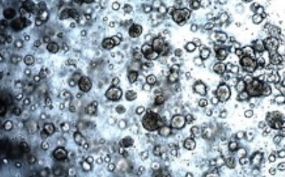
A) Acinar Lung PDO
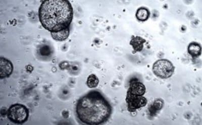
B) Mucinous Lung PDO
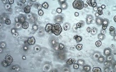
C) Solid Lung PDO
Figure 1 (A-C). Human Lung Cancer Organoids. Lung cancer PDOs derived from various NSCLC lung adenocarcinoma tumor biopsies have distinct morphologies when cultured in 3D cell cultures.
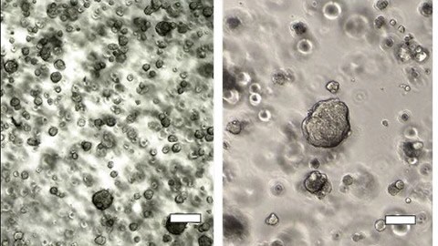
ORGANOIDS IN MATRIGEL
XDO.137 organoics passage 6.
Scale bar = 100µm
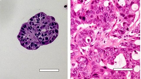
HISTOLOGY (HEMATOXYLIN & EOSIN)
XDO.137 organoics (left) and tissue of origin (right)
Scale bar = 100µm
Figure 2. Lung Cancer Organoid Cultured in 3D vs Native Tissue. Morphological and histological comparison of Acinar/Papillary 3dGRO™ Human Lung Organoids (XDO.137) (SCC604) cultured in 3D, passage 6 (Left) vs. native tissue of origin (Right).
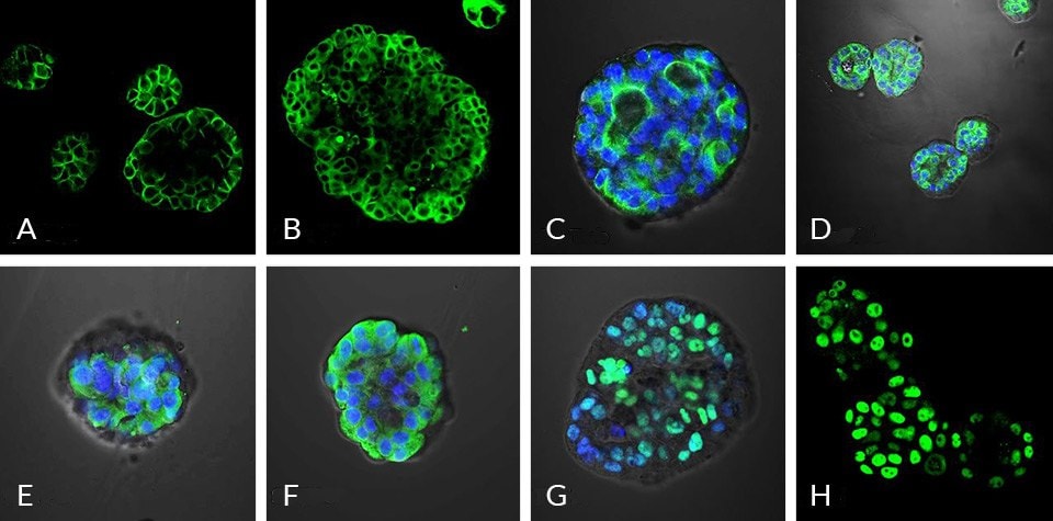
Figure 3. Immunocytochemical (ICC) Characterization of Human Lung Organoids.Lung cancer organoids are positive for (A) EPCAM, (B) pan-Cytokeratin, (C) acetylated α-tubulin, (D) Muc5AC, (E) surfactant protein-B (SFTPB), (F) surfactant protein-C (SFTPC), (G) TTF1 and (H) P63 protein expression.
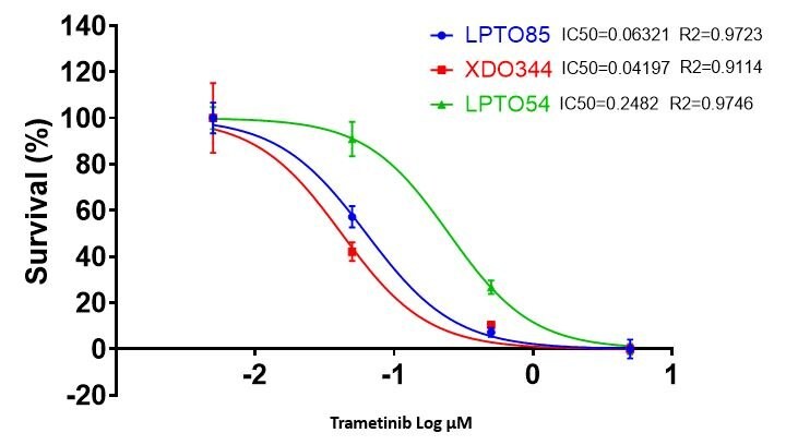
Figure 4. Drug Cytotoxicity Screening using Human Lung Cancer Organoids.RAS mutations have been found in approximately 30% of all human cancers, with KRAS as the most commonly mutated family member. Lung cancer organoid cytotoxicity was measured after exposing various NSCLC organoids (104 cells/well) to the MEK inhibitor Trametinib. The result shows KRAS mutant XDO344 and LPTO85 were much more sensitive to Trametinib (XDO344: IC50 0.04uM, LPTO85: IC50 0.06uM) than wild-type KRAS LPTO54 (LPTO54: IC50 0.25uM).
References

To continue reading please sign in or create an account.
Don't Have An Account?