Paper-based Sensing Devices for Point-of- care Testing: Current Advances
Introduction
The development of sensing platforms for promoting rapid, inexpensive, and accurate clinical diagnostics that can be used in point-of-care testing (POCT) has received noticeable attention in recent years.1 A wide range of devices designed in varied materials have been reported in the literature for multiple purposes associated with clinical screening and detection of target analytes. Among them, cellulosic paper-based substrates have emerged as simple, biocompatible, affordable, and easy-to-use platforms for POCT applications. The attractive features of this material make it a potential candidate for the development of commercial POCT devices, as it is already seen for pregnancy, Dengue, and COVID-19 diagnostic kits, which can be found in drug stores worldwide.2,3
The increasing popularity of paper platforms for POCT applications can be attributed to the inherent advantages of cellulosic material. Due to the paper porosity, the fluid can be handled spontaneously by capillary action, eliminating the need for external instruments for fluid pumping. In addition, paper is commercially available in different formats, ranging from pore size to substrate thickness, which directly affects flow rate. Knowledge of substrate properties is essential for a rational selection of the best material for the desired application since they play a crucial role in analytical performance.4
Paper-based platforms can be manufactured through different fabrication protocols, promoting the creation of microfluidic channels interconnected with detection zones for colorimetric readouts or conductive tracks for electrochemical sensing. Colorimetric and electrochemical detection are the most popular detectors associated with paper-based devices. Colorimetric detection can be performed by the naked eye, allowing rapid yes- or-no responses, which greatly appeals to POCT applications. However, the colorimetric readout is quantified by extracting the color intensity of a digitalized image using specific software or free applications (Apps). This opens the possibility of exploring portable scanners or smartphones for supporting quantitative analysis when required. In the same way, electrochemical measurement may be done using portable and battery-powered potentiostats that are compatible with smartphones and tablets, making their use possible in the POCT.1,5
Considering the immense potential of paper-based sensing devices for POCT and commercialization,4 this mini-review summarizes the up-to-the-minute advances in clinical diagnostics. A summary of the most common protocols for fabricating paper-based sensing platforms and very recent examples of clinical diagnostics are successfully presented and discussed.
Fabrication Techniques of Paper-based Sensors and Biosensors
Various fabrication methods of colorimetric or electrochemical paper-based analytical devices (PADs) for POCT are displayed in Figure 1.
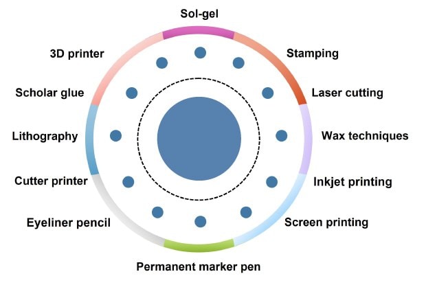
Figure 1.Summary of the most common techniques for the fabrication of paper-based analytical devices. Reproduced with permission from reference 26, copyright Springer 2014.
The colorimetric PADs include lateral flow assays (LFAs) and microfluidic PADs (μPADs). The formation of a hydrophobic boundary in μPADs is crucial to attaining a physical barrier for the sample distribution pathway toward detection zones. Its popularity is associated with the mass production of highly reproducible devices. The most common procedures to fabricate physical barriers are those based on wax techniques, such as wax printing5–8 and inkjet.9 In wax printing, a solid ink printer is used to delimit hydrophobic barriers on the paper’s surface. Applying hot lamination increases the wax’s permeability in the paper’s pores, improving insulation. Afterward, the reaction zone is impregnated with specific colorimetric reagents (chromogens) to interact with the target present in the sample, thus producing changes in the coloration.
In the case of LFA, the test and control lines are prepared using the streptavidin-biotin interaction10–12 principle to promote the conjugation with the recognition element that will further interact with the target.
Regarding electrochemical paper-based analytical device (ePAD) fabrication, the techniques most commonly used to manufacture ePADs are wax printing, screen printing, and stencil printing.13–15 Filter paper and Whatman® chromatography paper substrates are commonly used due to their versatility, porosity, flexibility, and robustness. Screen printing and stencil printing are employed to fabricate electrochemical biosensor electrodes. Conductive ink is applied to the substrate by squeezing it through a fine mesh screen or stencil mold, printing the paper surface.16 These technologies allow the mass production of cost-effective electrodes, enabling system miniaturization and reducing the sample volume for analysis. Automated production minimizes the issues related to the lack of reproducibility.
Materials and Practical Applications
Several ink formulations can be proposed, considering different solvent, binder, and conductive compound compositions for each specific application. Silva-Neto and collaborators17 created conductive ink using graphite flakes and waste acrylonitrile- butadiene-styrene (ABS) filaments used in 3D printing. The conductive ink was used in the confection of electrodes by stencil printing to make a paper-based electrochemical sensor to analyze S-nitroso-cysteine.
However, there are other methods of making even simpler electrodes. For instance, Souza et al.18 reported using graphite ink and nail polish on filter paper. Despite the easy and low-cost production of this electrode, large-scale manufacturing may be impractical due to the non-uniform application of conductive ink with a brush. However, to solve this problem, the authors polished the electrode and observed an increase in sensitivity.
With the electrodes manufactured, the next step is to modify the surface of the working electrode through electrodeposition, drop casting, and dipping with elements of target recognition, mediators, and receptors. This procedure is critical to improving the selectivity and sensitivity of the biosensor for biomarkers of clinical interest.19 For modification, compounds responsible for sample recognition and improvement of electron transfer are used, such as enzymes, aptamers, peptide nucleic acids, metallic nanoparticles, nanocomposites, monoclonal antibodies, metallic oxides, carbon nanotubes (Cat. Nos. 901046, 901019, 901634), polymers, antigens, Nafion™, carbon dots (Cat. Nos. 900726, 900713), etc.8,15,20
Gupta and Ghrera,20 synthesized a nanocomposite capable of transforming paper into a conductor to manufacture electrodes. The paper was dipped in a mix of polystyrene sulfonate (PEDOT:PSS, Cat. Nos. 483028, 900208, 900181) and gold nanoparticles and ultrasonicated for 40 min, promoting the deposition of PEDOT:PSS-AuNP on the cellulose fibers. Then, for the functionalization of the electrode to detect the biomarker procalcitonin, a monoclonal antibody (anti-PCT) and a blocking agent, BSA, were dropped over the working electrode. When anti- PCT and BSA were added, the peak current decreased due to the insulating characteristic of BSA, which reduced the transfer of electrons between the electrode-solution interface.
Due to the global COVID-19 outbreak, there was a need to develop devices capable of detecting the virus and assisting in monitoring and identifying infected people. Based on this, many efforts have been devoted to the development of ePADs capable of detecting SARS-CoV-2 using different bioreceptors, such as pyrrolidinyl peptide nucleic acid (acpcPNA), monoclonal antibody (mAb) CR3022 and complementary DNA (cDNA).22–24
For the fabrication of a DNA biosensor for COVID-19, Lomae et al.25 first demonstrated cellulose oxidation using a periodate solution, which resulted in the formation of aldehyde groups on the paper surface. Next, they synthesized an acpcPNA capable of binding to the SARS-CoV-2 cDNA. The ePAD functionalization occurred after a solution with acpcPNA and NaBH3CN in dimethylformamide (DMF, Cat. No. 5.89565) was dropped over the aldehyde-modified paper, promoting the immobilization of acpcPNA via covalent bonding.
POCT Applications Using Paper-based Devices
In recent years, POCT devices have emerged as powerful platforms for clinical analysis.27 Some advantages inherent to using such analytical devices are portability, ease of use, disposability (often environmentally friendly), rapid responses, accuracy, sensitivity comparable to more sophisticated techniques, and affordable costs. Additionally, because patients can frequently explore POCT platforms, they can help decentralize tests that are routinely conducted in hospitals and specialty clinics. In this sense, clinical analysis at the point of care plays a crucial role in assisting in the control of disease in the event of an epidemic or pandemic and in promoting early diagnosis and treatment.27,28
COVID-19 Diagnosis
Coronavirus disease 2019 (COVID-19) is caused by the severe acute respiratory syndrome coronavirus 2 (SARS-CoV-2) virus. Symptoms related to the SARS-CoV-2 infection range from cough and fever in simpler cases to more severe cases that can progress to pneumonia and death. One of the crucial characteristics of SARS-CoV-2 is its high level of transmissibility, which culminated in late 2019 in a pandemic that resulted in the deaths of more than 6 million people to date.29 Therefore, the rapid discovery of the SARS-CoV-2 infection is extremely important to initiate prompt treatment and prevent more people from being contaminated.
Regarding COVID-19 diagnosis, Reverse Transcriptase Polymerase Chain Reaction (RT-PCR) is considered a gold standard technique and presents the lowest limits of detection (LODs) and high specificity for results. However, RT-PCR is classified as a high-cost technology. On the other hand, employing analytical platforms for SARS-CoV-2 detection at the point-of-care proves to be an efficient and cost-effective approach. It enables rapid delivery of results, even in the initial stages of infection, including in asymptomatic patients. Some common examples of COVID-19 diagnostic devices developed considering paper platforms in 2023 are lateral flow immunoassay devices, colorimetric sensors (spot-test or microfluidics), and electrochemical devices. Some examples of these are mentioned in the next sections.28
Lateral Flow Immunoassay Devices
Recently, Lee et al.30 developed a colorimetric lateral flow immunoassay (LFIA) based on oriented antibody immobilization for rapid, accurate, sensitive, and early detection of SARS-CoV-2 trough receptor binding domain (RBD) (See Figure 2). Some materials, such as cellulose membrane and absorbent pads, were successfully used for fabricating the analytical device. For this purpose, a CBP31-BC bio-functional linker was used to immobilize antibody-conjugated gold nanoparticles (AuNPs, Cat. Nos. 928070, 928186, 927856, and 928194) on a cellulose membrane, recognizing the antigens to form antigen-antibody complexes—the proposed LFIA presents visual detection of SARS-CoV-2 in 15 min. The relationship of RBD concentrations ranging from 0.05 to 200 ng mL-1 was analyzed. The appreciable limit of detection (LOD) was estimated to be equal to 5 × 10⁴ copies mL-1.
To verify the applicability of the device, 19 real nasopharyngeal samples were analyzed along side healthy samples, then compared to RT-PCR results, which demonstrated 100% accuracy (including samples with low viral loads). The results presented the proposed analytical platform as a rapid and accurate strategy to promote point-of-care testing to control COVID-19 proliferation.
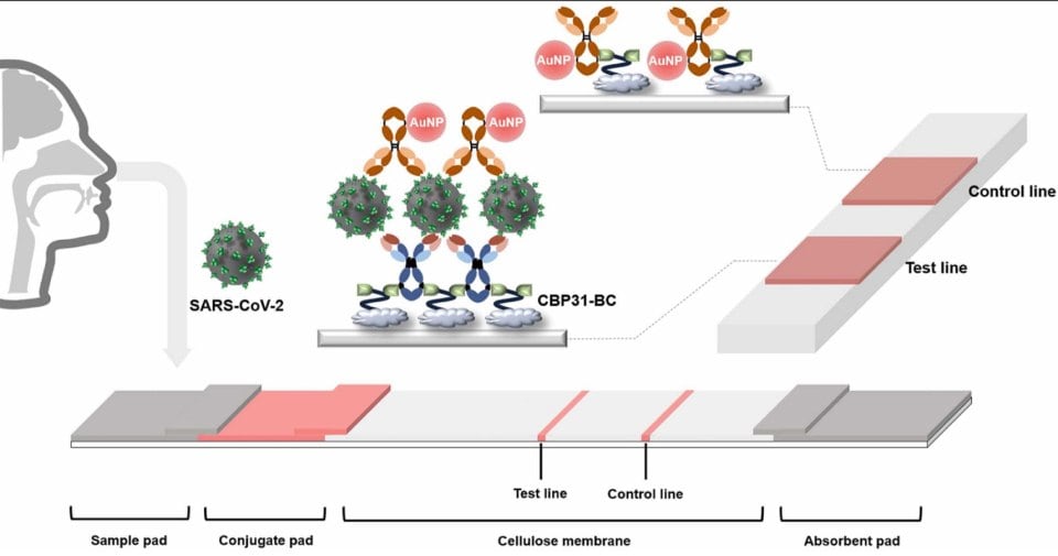
Figure 2.Schematic illustration of the CBP31-BC-based LFIA for detecting SARS-CoV-2. Reproduced with permission from reference 30, copyright Elsevier 2023.
Microfluidic Devices
Recently, Li et al.9 successfully proposed a rotational paper-based sensing platform coupled with a tetrahedral DNA framework (TDF) for discriminating between SARS-CoV-2 and influenza A H1N1 virus (Figure 3 A–C). Regarding the construction of the device, Whatman filter paper was wax-printed and heated to create the hydrophobic barrier. Posteriorly, the reaction between horseradish peroxidase (HRP) and 3,3′,5,5′-tetramethylbenzidine (TMB)–H2O2 was explored. Thus, the color change obtained by colorimetric response was analyzed in grayscale using free ImageJ software. Spike protein and H1N1 were detected with adequate sensitivity, considering the buffer and swab virus preservation solution matrix. Therefore, the portable, low-cost, and disposable device provided a reliable method for rapid clinical differentiation and diagnosis of COVID-19 and influenza A.
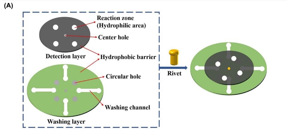

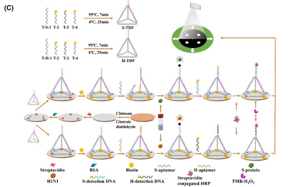
Figure 3.Microfluidic device to detect SARS-CoV-2. A) Graphical representation of paper analytical device. B) Schematic of the working process of analytical device composed by “detection/washing” controlled by rotating the washing layer. C) Analytical procedure for detecting of S protein and H1N1. Reproduced with permission from reference 25, copyright 2023 Elsevier.
Electrochemical Devices
Lomae et al.25 manufactured a novel disposable paper-based electrochemical sensor for detecting SARS-CoV-2 (Figure 4A) using Whatman chromatography paper as the substrate. For electrode construction, graphene conductive ink (Cat. Nos. 900450, 900695, 900960) and silver/silver chloride (Ag/AgCl, Cat. Nos. 901090, 923575, 907669, 923559, 923567) ink were explored. The strategy used consisted of a working electrode (WE) modification using pyrrolidinyl peptide nucleic acid (acpcPNA) as the biological recognition element to capture the target complementary DNA (cDNA). In this sense, the electrochemical response provided by a redox probe decreased in the presence of the target cDNA since hybridization with acpcPNA promotes a block of the WE surface. This way, the electrochemical readout was correlated to SARS-CoV-2 concentration. It is important to mention that the proposed method was explored using a portable smartphone-assisted Sensit Smart Potentiostat (PalmSens) to record data. The PNA-based electrochemical paper-based analytical device (PNA-based ePAD) is of high specificity toward the SARS-CoV-2 N gene due to highly selective PNA-DNA binding and satisfactory detection limit (1.0 pmol L−1). Finally, the PNA- based ePAD was successfully used to promote the detection of amplification-free SARS-CoV-2 in 10 nasopharyngeal swab samples (7 positive and 3 negative), providing results with 100% agreement with results recorded using a gold technique (RT-PCR).
Jaewjaroenwattana et al.23 proposed a simple and low-cost electroanalytical platform to promote SARS-CoV-2 antigen detection (Figure 4B). For fabricating the disposable paper- based immunosensor, a plant-based anti-SARS-CoV-2 monoclonal antibody CR3022, expressed in Nicotiana benthamiana, is a cost- effective approach. To achieve effective antibody immobilization, an endeavor was made to enhance the presence of COOH functional groups on the electrode surface. This was accomplished by modifying the screen-printed graphene electrode with cellulose nanocrystals. Differential pulse voltammetry was used for the electrochemical readout provided by the [Fe(CN)6]3-/4- redox probe, allowing quantification of the presence of the receptor binding domain (RBD) spike protein of SARS-CoV-2. The proposed method provided satisfactory performance, including linearity in the concentration range from 0.1 pg mL-1 to 500 ng mL-1 and a limit of detection of 2.0 fg mL-1. The feasibility of the electrochemical immunosensor was successfully tested for detecting RBD in nasopharyngeal swab samples that were in concordance when compared with RT-PCR analysis. Moreover, different levels of RBD in spiked artificial saliva samples were tested, showing the ability of the sensor to detect the target analyte in different matrices and highlighting the potential use of the paper-based electrochemical sensor as a POCT device for COVID-19 diagnosis.
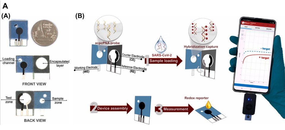

Figure 4.Electrochemical detection of SARS-CoV-2. A1) Schematic representation of PNA-based ePAD. A2) Representation of electroanalytical measurement using a smartphone-based potentiostat. Reproduced with permission from reference 9, copyright 2023 Elsevier. B) Design and the detection procedure using paper-based electrochemical immunosensor dedicated to detection of SARS-CoV-2. Reproduced with permission from reference 23, copyright 2023 Elsevier.
Conclusion and Perspectives
This review provided an overview of the pocket-sized point-of-care technologies that can open many possibilities for healthcare applications with the promise of inexpensive, fast, and precise analyses. Some examples of materials employed to construct paper devices were reviewed. Chromatographic paper, carbon allotropes, gold nanoparticles, and specific antibodies are primarily suitable for biological sensing applications. Many aspects of paper-based sensors need to be developed for further progress in the field, such as the full validation of clinical trials through large-scale investigations with the fundamental collaboration of medical professionals. Considering their versatility, these devices require significant work to advance beyond prototypes into commercial use. We believe the proposed paper can be routinely employed to increase the use of paper-based devices for point-of-care testing.
Acknowledgments
The authors would like to thank CAPES (finance code 88887.192880/2018-00), CNPq (grants 307554/2020-1, 401256/2020-0 and 405620/2021-7), and INCTBio (grant 465389/2014-7) for the financial support and granted scholarships.
References
如要继续阅读,请登录或创建帐户。
暂无帐户?