Cleaning Up Samples using 2-D Clean-Up Kit
2-D Clean-Up Kit is designed to prepare samples that would otherwise produce poor 2-D results due to high conductivity, high levels of interfering substances, or low concentration of protein.
Current methods of protein precipitation suffer from several significant disadvantages:
- Precipitation can be incomplete, resulting in the loss of proteins from the sample and introduction of bias into the 2-D result.
- The precipitated protein can be difficult to resuspend and often cannot be fully recovered.
- The precipitation procedure can itself introduce ions that interfere with first-dimension IEF.
- Precipitation can be time-consuming, requiring overnight incubation of the sample.
2-D Clean-Up Kit circumvents these disadvantages by providing a method for selectively precipitating protein for 2-D electrophoresis. Protein can be quantitatively precipitated from a variety of sources without interference from detergents, chaotropes, and other common reagents used to solubilize protein. Recovery is generally greater than 90%. The procedure does not result in spot gain or loss, or changes in spot position relative to untreated samples. The precipitated proteins are easily resuspended in 2-D sample solution. The procedure can be completed in less than one hour.
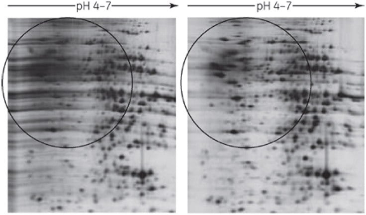
The overall quality of protein separation using 2-D Clean-Up Kit has been shown to be superior to that of samples prepared by precipitation with acetone (54). Preparation of protein samples with the kit reduces horizontal streaking, improves spot resolution, and increases the number of spots detected compared with samples treated by other means (Figure 1 and Table 1).

Figure 1. 2-D Clean-Up Kit eliminates horizontal streaking caused by residual SDS. Sample: Rat liver extracted with 4% SDS, 40 mM Tris base. First dimension: Approximately 20 μg rat liver protein, 7-cm Immobiline DryStrip pH 4–7, Ettan IPGphor Isoelectric Focusing System 17.5 kVh. Second dimension: SDS-PAGE (12.5%), SE 260 (8 × 9 cm gel). Stain: Silver Staining Kit, Protein.
* Protein spots were detected using ImageMaster™ 2D Elite software.
† 9.8 M urea, 2% CHAPS, 0.5% IPG Buffer pH 3–10, 65 mM DTT.
The 2-D Clean-Up Kit procedure uses a combination of a unique precipitant and co-precipitant to quantitatively precipitate the sample proteins while leaving interfering substances behind in the solution. The proteins are pelleted by centrifugation and the precipitate is washed to further remove non-protein contaminants. The mixture is centrifuged again and the resultant pellet can be easily resuspended into a 2-D sample solution of choice, compatible with first dimension IEF.
The kit contains sufficient reagents to process 50 samples of up to 100 μl each. The procedure can be scaled-up for larger volumes or more dilute samples.
Protocol: 2-D Clean-Up Kit
Reagents supplied
Precipitant, co-precipitant, wash buffer, wash additive.
Required but not provided
Ice bath, 1.5-mL capped microcentrifuge tubes, microcentrifuge capable of at least 12 000 × g, rehydration solution or IEF sample solution for resuspension (see next section), vortex mixer.
Preliminary notes
Procedure A is applicable for sample volumes of 1–100 μl containing 1–100 μg of protein. For larger samples containing more than 100 μg of protein, use procedure B.
Prior to starting the procedure, chill the wash buffer to -20 °C for at least 1 h.
Procedure A: For sample volumes of 1–100 μl (containing 1–100 μg of protein per sample)
Process the protein samples in 1.5-mL microcentrifuge tubes. All steps should be carried out on ice unless otherwise specified.
- Transfer 1–100 μl of protein sample (containing 1–100 μg protein) into a 1.5-mL microcentrifuge tube.
- Add 300 μl of precipitant. Mix well by vortexing or inversion. Incubate the tube on ice (4–5 °C) for 15 min.
- Add 300 μl of co-precipitant to the mixture of protein and precipitant. Mix by vortexing briefly.
- Position the tubes in a microcentrifuge with cap-hinges facing outward. Centrifuge at maximum speed (at least 12 000 × g) for 5 min. Remove the tubes from the microcentrifuge as soon as centrifugation has finished. A small pellet should be visible. Proceed rapidly to the next step to avoid resuspension or dispersion of the pellet.
- Remove as much of the supernatant as possible by decanting or careful pipetting. Do not disturb the pellet.
- Carefully reposition the tubes in the microcentrifuge with the cap-hinges and pellets facing outward. Centrifuge the tubes briefly to bring any remaining liquid to the bottom of the tubes. Use a pipette to remove the remaining supernatant. There should be no visible liquid remaining in the tubes.
- Without disturbing the pellet, layer 40 μl of co-precipitant on top of each pellet. Incubate the tubes on ice for 5 min.
- Carefully reposition the tubes in the centrifuge with the cap-hinges facing outward. Centrifuge for 5 min. Use a pipette to remove the supernatant.
- Pipette 25 μl of distilled or deionized water on top of each pellet. Vortex each tube for 5–10 s. The pellet should disperse, but not dissolve in the water.
- Add 1 mL of wash buffer (prechilled for at least 1 h at -20 ºC) and 5 μl of wash additive to each tube. Vortex until the pellets are fully dispersed.
Note: The protein pellet will not dissolve in the wash buffer. - Incubate the tubes at -20 °C for at least 30 min. Vortex for 20–30 s once every 10 min. At this stage, the tubes can be stored at -20 ºC for up to one week with minimal protein degradation or modification.
- Centrifuge the tubes at maximum speed (at least 12 000 × g) for 5 min.
- Carefully remove and discard the supernatant. A white pellet should be visible. Allow the pellet to air dry briefly (no more than 5 min).
Do not overdry the pellet. If it becomes too dry, it will be difficult to resuspend. - Resuspend each pellet in an appropriate volume of rehydration or IEF sample loading solution for first-dimension IEF. See next section for examples of rehydration solutions and volumes appropriate to different applications. Vortex the tubes for at least 30 s. Incubate at room temperature. Vortex or aspirate and dispense using a pipette to fully dissolve.
If the pellet is large or too dry, it may be difficult to resuspend fully. Sonication or treatment with the Sample Grinding Kit can speed resuspension. - Centrifuge the tubes at maximum speed (at least 12 000 × g) for 5 min to remove any insoluble material and to reduce any foam. The supernatant may be loaded directly onto first-dimension IEF or transferred to another tube and stored at -80 ºC for later analysis.
Procedure B: For larger samples of more than 100 μg of protein
All steps should be carried out on ice unless otherwise specified.
- Transfer the protein samples into tubes that can be centrifuged at 8000 × g. Each tube must have a capacity at least 12-fold greater than the volume of the sample. Use only polypropylene, polyallomer, or glass tubes.
The wash buffer used later in the procedure is not compatible with many plastics. This limits the choice of centrifuge tube materials. - For each volume of sample, add three volumes of precipitant. Mix well by vortexing or inversion. Incubate on ice (4–5 °C) for 15 min.
- For each original volume of sample, add three volumes of co-precipitant to the mixture of protein and precipitant. Mix by vortexing briefly.
- Position the tubes in a microcentrifuge with the cap-hinges facing outward. Centrifuge at 8000 × g for 10 min. Remove the tubes from the microcentrifuge as soon as centrifugation has finished. A pellet should be visible.
Proceed rapidly to the next step to avoid resuspension or diffusion of the pellet. - Remove as much of the supernatant as possible by decanting or careful pipetting. Do not disturb the pellet.
- Carefully position the tubes in the microcentrifuge with the cap-hinges and pellets facing outward. Centrifuge the tubes for at least 1 min to bring any remaining liquid to the bottom of the tubes. Use a pipette to remove the remaining supernatant. There should be no visible liquid remaining in the tubes.
- To each tube, add threefold to four-fold more co-precipitant than the size of the pellet.
- Carefully reposition the tubes in the microcentrifuge with the cap-hinges facing outward. Centrifuge for 5 min. Use a pipette to remove the supernatant.
- Pipette enough distilled or deionized water on top of each pellet to cover the pellet. Vortex each tube for several seconds. The pellets should disperse, but not dissolve in the water.
- Add 1 mL of wash buffer, prechilled for at least 1 h at -20 ºC to each tube. (For an initial sample volume of 0.1–0.3 mL, add 1 mL of wash buffer. However, the volume of wash buffer must be at least 10-fold greater than the distilled/deionized water added in step 9.) Add 5 μl wash additive (use only 5 μl wash additive, regardless of the original sample volume). Vortex until the pellet is fully dispersed.
Note: The protein pellet will not dissolve in the wash buffer. - Incubate the tubes at -20 °C for at least 30 min. Vortex for 20–30 s once every 10 min. At this stage, the tubes can be stored at -20 ºC for up to one week with minimal protein degradation or modification.
- Centrifuge the tubes at 8000 × g for 10 min.
- Carefully remove and discard the supernatant. A white pellet should be visible. Allow the pellet to air dry briefly (no more than 5 min).
Do not over-dry the pellet. If it becomes too dry, it will be difficult to resuspend. - Resuspend each pellet in rehydration solution for first-dimension IEF. The volume of rehydration solution used can be as little as 1/20 of the volume of the original sample. See next section for examples of rehydration solutions and volumes appropriate for different applications. Vortex the tube for 30 s. Incubate at room temperature. Vortex or aspirate and dispense using a pipette to fully dissolve.
If the pellet is large or too dry, it may be difficult to resuspend fully. Sonication can speed resuspension. - Centrifuge the tubes at 8000 × g for 10 min to remove any insoluble material and to reduce any foam. The supernatant may be loaded directly onto first-dimension IEF or transferred to another tube and stored at -80 ºC for later analysis.
Resuspension of pellet
2-D Clean-Up Kit produces a protein pellet. When using cup loading, resuspend the pellet in sample preparation solution (see appendix I). When using rehydration loading, resuspend the pellet in rehydration solution (see options 1 and 2 below), which is applied directly to the Immobiline DryStrip gel.
- Rehydration solution containing 8 M urea
Use solution C in appendix I. This all-purpose solution gives clean, sharp 2-D separations. - Rehydration solution containing 7 M urea and 2 M thiourea
Use solution D in appendix I. This is a more strongly solubilizing solution that results in more spots in the final 2-D pattern.
Any other components added to the rehydration solution must either be uncharged or present at a concentration of less than 5 mM. The addition of salts, acids, bases, and buffers is not recommended. - DeStreak Reagent
Use for basic strips. See section 2.6.2 for details on the reagent.
Sample resuspension volumes
The volume of rehydration solution used to resuspend the sample depends on the sample loading method and the length of the Immobiline DryStrip gel used for the first-dimension separation. If using Ettan IPGphor 3 and the sample is to be loaded onto the Immobiline DryStrip gel using a sample cup, the sample volume should not exceed 150 μl. If the sample is to be loaded onto the Immobiline DryStrip gel by rehydration, the sample volumes shown in Table 2 should be used according to the length of the Immobiline DryStrip gel.
The optimal quantity of protein to load varies widely depending on factors such as sample complexity, the length and pH range of the Immobiline DryStrip gel, and the method of visualizing the 2-D gel separation. General guidelines are given in chapter 2.
The protein concentration of the sample is best determined using the 2-D Quant Kit, which can accurately quantitate protein in the presence of detergents, reductants, and other reagents used in sample preparation.
* DTT is added just prior to use: 7 mg DTT per 2.5-mL aliquot of rehydration stock solution. For rehydration loading, sample is also
added to the aliquot of rehydration solution just prior to use.
† If necessary, the concentration of urea can be increased to 9 M or 9.8 M.
‡ Other neutral or zwitterionic detergents may be used at concentrations up to 2% (w/v). Examples include Triton X-100, NP-40, octylglucoside, and the alkylamidosulfobetaine detergents ASB-14 and ASB-16 (Calbiochem).
§ As an alternative to IPG Buffer, use Pharmalyte 3–10 for Immobiline DryStrip 3–10 or 3–10 NL, Pharmalyte 5–8 for Immobiline
DryStrip 4–7.
¶ A Pharmalyte/IPG Buffer concentration of 0.5% (125 μl) is recommended with Ettan IPGphor 3 Isoelectric Focusing System and an IPG Buffer/Pharmalyte concentration of 2% (500 μl) is recommended with the Multiphor II and Immobiline DryStrip Kit system.
Store in 2.5-mL aliquots at -20 °C.
* DTT is added just prior to use: Add 7 mg DTT per 2.5-mL aliquot of rehydration stock solution.
† Other neutral or zwitterionic detergents may be used at concentrations up to 2% (w/v). Examples include Triton X-100, NP-40, octylglucoside, and the alkylamidosulfobetaine detergents ASB-14 and ASB-16 (Calbiochem).
‡ A Pharmalyte/IPG Buffer concentration of 0.5% (125 μL) is recommended with Ettan IPGphor 3 Isoelectric Focusing System and an IPG Buffer/Pharmalyte concentration of 2% (500 μL) is recommended with the Multiphor II and Immobiline DryStrip Kit system.
Store in 2.5-mL aliquots at -20 °C.
Materials
To continue reading please sign in or create an account.
Don't Have An Account?