
NA57
Anti-DNA-PK (Ab-2) Mouse mAb (18-2)
liquid, clone 18-2, Calbiochem®
别名:
Anti-DNA Dependent Protein Kinase
登录 查看组织和合同定价。
选择尺寸
关于此项目
NACRES:
NA.43
UNSPSC Code:
12352203
Clone:
18-2, monoclonal
Species reactivity:
human
Application:
—
Citations:
11
biological source
mouse
antibody form
purified antibody
antibody product type
primary antibodies
clone
18-2, monoclonal
form
liquid
contains
≤0.1% sodium azide as preservative
species reactivity
human
manufacturer/tradename
Calbiochem®
storage condition
OK to freeze
dilution
(Frozen Sections (1:50-1:100)
Immunoblotting (1:250-1:500)
Immunofluorescence (1:50-1:100)
Immunoprecipitation (10 µL/mg protein lysate)
Neutralization Studies
Paraffin Sections (1:50-1:100, heat/pressure cooker pre-treatment required))
isotype
IgG1
shipped in
wet ice
Quality Level
Gene Information
human ... PRKDC(5591)
target post-translational modification
unmodified
General description
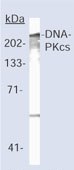
Purified mouse monoclonal antibody generated by immunizing BALB/c mice with the specified immunogen and fusing the splenocytes with mouse myeloma SP2/0 cells (see application references). Recognizes the ~460 kDa DNA-PK protein.
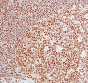
Recognizes the ~470 kDa DNA-PK protein in A431, HeLa, and HL60 cells, normal tonsil, normal colon, and breast carcinoma tissue.
This Anti-DNA-PK (Ab-2) Mouse mAb (18-2) is validated for use in Frozen Sections, Immunoblotting, IF, IP, Neutralization Studies, Paraffin Sections for the detection of DNA-PK (Ab-2).
Immunogen
DNA-PK purified from HeLa cells
Epitope: within amino acids 1-2713 of DNA-PK
Human
Application
Frozen Sections (1:50-1:100)
Immunoblotting (1:250-1:500)
Immunofluorescence (1:50-1:100)
Immunoprecipitation (10 l/mg protein lysate)
Neutralization Studies (see comments)
Paraffin Sections (1:50-1:100, heat/pressure cooker pre-treatment required)
Packaging
Please refer to vial label for lot-specific concentration.
Physical form
In 10 mM PBS, 0.2% BSA, pH 7.4.
Preparation Note
For long-term storage, aliquot and freeze (-20°C). Avoid freeze/thaw cycles.
Analysis Note
Positive Control
A-431, HeLa, or HL-60 cells or normal tonsil, normal colon, or breast carcinoma tissue
A-431, HeLa, or HL-60 cells or normal tonsil, normal colon, or breast carcinoma tissue
Other Notes
Kharbanda, S., et al. 1997. Nature386, 732.
McConnell, K.R., et al. 1997 J. Immunol.158, 2083-2089.
Peterson, S.R., et al. 1997. J. Biol. Chem.272, 10227.
Anderson, C.W. and Carter, T.H. 1996. Curr. Top. Microbiol. Immunol.217, 91.
Blunt, T., et al. 1996. Proc. Natl. Acad. Sci. USA93, 10285.
Carpenter, C.L. and Cantley, L.C. 1996. Curr. Opin. Cell. Biol.8, 153.
Chan, D.W. and Lees-Miller, S.P. 1996. J. Biol. Chem.271, 8936.
Connelly, M.A., et al. 1996. Gene175, 271.
Danska, J.S., et al. 1996. Mol. Cell. Biol.16, 5507.
Han, Z., et al. 1996. J. Biol. Chem.271, 25035.
Lees-Miller, S.P. 1996. Biochem. Cell. Biol.74, 503.
Song, Q., et al. 1996. EMBO J.15, 3238.
Teraoka, H., et al. 1996. FEBS Lett.393, 1.
Blunt, T., et al. 1995. Cell80, 813.
Hartley, K.O., et al. 1995. Cell82, 849.
Kirchgessner, C.U., et al. 1995. Science267, 1178.
Lees-Miller, S.P., et al. 1995. Science267, 1183.
Poltoratsky, V.P., et al. 1995. J. Immunol.155, 4529.
Anderson, C.W. 1993. Trends Biochem. Sci.18, 433.
Gottleib, T.M. and Jackson, S.P. 1993. Cell72, 131.
This antibody will inhibit (approximately 50%) the kinase activity of DNA-PKcs. However, preincubation of the enzyme with DNA protects DNA-PKcs against inactivation by Clone 18-2. Antibody should be titrated for optimal results in individual systems.
Legal Information
CALBIOCHEM is a registered trademark of Merck KGaA, Darmstadt, Germany
Disclaimer
Toxicity: Standard Handling (A)
未找到合适的产品?
试试我们的产品选型工具.
存储类别
12 - Non Combustible Liquids
wgk
WGK 2
flash_point_f
Not applicable
flash_point_c
Not applicable
Tewodros Mamo et al.
Biochemical and biophysical research communications, 486(2), 307-313 (2017-03-17)
Osteosarcoma survival rate has not improved over the past three decades, and the debilitating side effects of the surgical treatment suggest the need for alternative local control approaches. Radiotherapy is largely ineffective in osteosarcoma, indicating a potential role for radiosensitizers.
Xiaosheng Wu et al.
Journal of immunology (Baltimore, Md. : 1950), 174(2), 934-941 (2005-01-07)
Activation-induced cytidine deaminase (AID) is required for Ig class switch recombination, a process that introduces DNA double-strand breaks in B cells. We show in this study that AID associates with the DNA-dependent protein kinase catalytic subunit (DNA-PKcs) promoting cell survival
Benu Brata Das et al.
The EMBO journal, 28(23), 3667-3680 (2009-10-24)
Human tyrosyl-DNA phosphodiesterase (TDP1) hydrolyzes the phosphodiester bond at a DNA 3' end linked to a tyrosyl moiety. This type of linkage is found at stalled topoisomerase I (Top1)-DNA covalent complexes, and TDP1 has been implicated in the repair of
Stéphanie Solier et al.
Proceedings of the National Academy of Sciences of the United States of America, 109(32), 12866-12872 (2012-07-04)
The "apoptotic ring" is characterized by the phosphorylation of histone H2AX at serine 139 (γ-H2AX) by DNA-dependent protein kinase (DNA-PK). The γ-H2AX apoptotic ring differs from the nuclear foci patterns observed in response to DNA-damaging agents. It contains phosphorylated DNA
Kristen E Hurov et al.
Genes & development, 24(17), 1939-1950 (2010-09-03)
In response to DNA damage, cells activate a complex signal transduction network called the DNA damage response (DDR). To enhance our current understanding of the DDR network, we performed a genome-wide RNAi screen to identify genes required for resistance to
我们的科学家团队拥有各种研究领域经验,包括生命科学、材料科学、化学合成、色谱、分析及许多其他领域.
联系客户支持