Cultrex® 3-D Culture Matrix™ Laminin I Protocol
Product Number 3446-005-01
I. Product Description
3D Culture is a cell culture method that provides cells with the necessary structure and signaling cues to direct reconstruction of the tissue architecture, and these methods provide physiologically predictive in vitro models for evaluating development and disease. Normal cells assemble into organoids that structurally resemble their emanating tissues, exhibit a polarized morphology, undergo cell cycle regulation, and produce tissue specific proteins1-6. While cancer cells assemble into tumor-like structures, lacking an organized architecture or cell cycle regulation and exhibiting tumor-specific markers depending on the extend of malignancy7-9. To aid in the advancement of this technology, we present Cultrex® 3-D Culture Matrix™ RGF BME which is the first Basement Membrane Extract produced and qualified specifically for use in 3D culture studies. The 3-D Culture Matrix™ RGF BME provides the foundation for cells to grow in three dimensions allowing for the formation of structures in vitro. To provide the most standardized basement membrane extract for use in 3D cultures, a special process is employed to reduce growth factors and manufacture matrix at a standard concentration of approximately 15 mg/mL. This product is then evaluated in a 3D culture to validate efficacy.
II. Specifications
Concentration: Please refer to lot specific data
Purity: ≥90% (SDS-PAGE)
Source: Murine Engelbreth-Holm-Swarm (EHS) tumor
Storage buffer: Dulbecco’s Modified Eagle’s medium without phenol red (Product No. D5030), with 10 μg/mL gentamicin sulfate (Product No. G1264)
III. Precautions and Limitations
For Research Use Only. Not for use in diagnostic procedures.
The physical, chemical, and toxicological properties of these products may not yet have been fully investigated; therefore, we recommend the use of gloves, lab coats, and eye protection while using these chemical reagents.
IV. Material Qualification
1. Functional Assays
Cell Attachment – Tested for the ability to promote cell attachment and spreading of MG63 human osteosarcoma cells.
3D culture – Laminin I promotes attachment and growth of a human epithelial cell line derived from mammary gland (MCF-10A) and human prostate (PC-3), and in the presence of assay medium, these cell lines differentiate into acinar structures.
2. Sterility Testing
No bacterial or fungal growth detected after incubation at 37 ⁰C for 14 days following USP XXIV Chapter 71 sterility test
No mycoplasma contamination detected by PCR
Endotoxin concentration ≤20 EU/mL by LAL assay
3. Gelling
1. Laminin I forms a firm gel at neutral pH and 37 ⁰C at 6 mg/mL.
V. Storage and Stability
Store at – 20 ⁰C in a manual defrost freezer or at – 80 ⁰C.
VI. 3-D Culture Protocol
This procedure must be conducted in an aseptic environment, such as a laminar flow hood or clean room, using aseptic technique to prevent contamination.
- Culture cells as recommended by cell supplier to establish a stable population at 37 ⁰C in a CO2 incubator; growth media, growth factors, serum requirements, and incubation period may vary by cell type, e.g. MCF-10A (DMEM, 5% Horse Serum, 20 ng/mL hEGF, 500 ng/mL Hydrocortisone, 100 ng/mL Cholera Toxin, 10 μg/mL Insulin, 1X Pen/Strep) and PC-3 (RPMI, 10% Horse Serum, 5% Fetal Bovine Serum).
- Thaw 3-D Culture Matrix Laminin I at 4 ⁰C overnight.
- Working on ice, add 250 μL of 3-D Culture Matrix Laminin I to each well in a sterile 48 well plate (enough matrix is supplied to assay approximately 20 wells); incubate plate at 37 ⁰C overnight to promote gelling of matrix.
- Working on ice, add 98 mL of growth media (as recommended by cell supplier) and 2 mL of 3-D Culture Matrix Laminin I or other differentiation factor (final concentration of 2%) to a sterile container, and label this container “Assay Media,” and swirl to mix. Any unused Laminin I can be stored at 4 ⁰C up to one week or stored in working aliquots at –20 ⁰C in a manual defrost freezer.
- Incubate Assay Medium at 37 ⁰C for 30 minutes in preparation for cell dilution.
- Harvest cells from culture, and dilute cells to 1 x 10⁴ cells/mL in 25 mL (total volume) of Assay Medium.
- Add 500 μL of cell suspension to each well of the 48 well plate containing 3-D Culture Matrix Laminin I (5,000 cells/well).
- Incubate plate at 37 ⁰C in a CO2 incubator overnight.
- Each day, observe cell growth and structure formation via inverted microscope.
- On day 4, carefully pipette off old media using a sterile serological pipette, and replace with new Assay Medium. Repeat on day 8 and day 12.
- When structures have grown to desired size, prepare cells for analysis (as recommended by manufacturer), and analyze structures. This point is dependent on cell line and growth conditions. In our qualification, MCF-10A cells are analyzed at 16 days, and PC-3 cells are analyzed at 10 to 12 days
Recommendations for analysis
- To fix cells, incubate for 20 minutes in 2% formalin, 1X PBS at room temperature.
- Cells may be analyzed in the plate on Laminin I; they may be transferred to a microscope slide (very carefully); or they may be embedded in paraffin and sectioned.
- Cells may also be isolated from the Laminin I and processed for protein, DNA or RNA analysis.

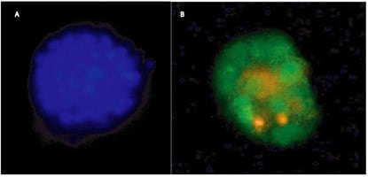
3-D Culture of MCF-10A Breast Cancer Cells using 3-D Culture Matrix™ Laminin I for 15 days in the presence of 2% Liminin I in Assay Medium, and stained using A) CPA Dye 2, and B) SYBR® Green (nuclear) and propidium iodine (dead or necrotic).
Materials
如要继续阅读,请登录或创建帐户。
暂无帐户?