Vascular Pericytes: Frequently Asked Questions (FAQs)

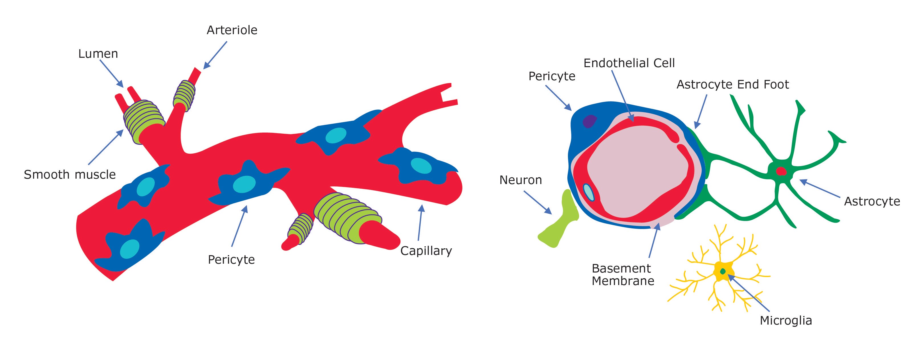
Figure 1.Vascular pericyte overview. Pericytes surround blood vessel capillaries and endothelial cells, neurons and astrocytes within the blood-brain-barrier (BBB) neurovascular unit.
Where are pericytes located?
Pericytes wrap around endothelial cells and are located within the basement membrane of capillary and postcapillary blood vessels. Pericytes are also key components of the neurovascular unit including endothelial cells, astrocytes, and neurons and maintain proper blood–brain-barrier (BBB) function in the CNS.
How do pericytes regulate angiogenesis?
Pericytes can produce vasoconstriction and vasodilation of capillaries to regulate vascular diameter and capillary blood flow. During angiogenesis, vascular pericytes are recruited by endothelial cells via platelet-derived growth factor (PDGF)-B/PDGF receptor-β (PDGFR-β) interactions. Pericytes are also associated with the regulation of vessel permeability, endothelial cell proliferation, and even leukocyte recruitment.
Are pericytes involved in cancer?
The analysis of tumor angiogenesis in experimental models of cancer has demonstrated that tumors have an immature vascular phenotype with continuous microvessel growth and remodelling. High pericyte densities within tumors have also been associated with forms of cancers that are the most aggressive and refractory to therapy. It is also thought that some pericytes are invloved in cancer EMT transitions (pericyte-fibroblast transition (PFT)) and are involved in regulating the tumor microenvironment. Vascular pericytes significantly contribute to tumor invasion and metastasis and are targets of new cancer therapies.
How do pericytes regulate blood-brain barrier function?
Pericytes, along with neurons, astrocytes, microglia, and endothelial cells comprise the neurovascular unit of the BBB. Blood -brain barrier permeability occurs by endothelial transcytosis regulated by pericytes by at least two mechanisms: 1) by regulating BBB-specific gene expression patterns in endothelial cells and 2) by inducing polarization of astrocyte end-feet surrounding CNS blood vessels.
Are pericytes involved in Alzheimer’s disease?
Reduced cerebral blood flow and vasculature resistance in capillaries occurs early in Alzheimer's disease (AD) and other dementias. Pericyte loss leads to Alzheimer-like symptoms in laboratory animals with elevated brain Aβ40 and Aβ42 levels and accelerates amyloid angiopathy, tau pathology and cerebral β-amyloidosis. Thus, pericytes may represent a new novel therapeutic target to modify disease progression in AD. Recent evidence suggests that brain pericytes modulate neuroinflammation by generating microglia phenotypes after injury.
Are all percicytes mesenchymal stem cells (MSCs)?
Mesenchymal stem cells (MSCs) are multipotent progenitors cells isolated from bone marrow, adipose and dental pulp. Both pericytes and mesenchymal stem cells can differentiate into osteocytes, adipocytes and chondrocytes and often express the same surface markers. MSCs have also been found in the perivascular space in virtually all organs. However, the precise identity of MSCs within the vascular wall is highly controversial. Recent evidence may suggest that perivascular MSCs act as precursors of pericytes and other stromal cells during tissue homeostasis
What markers are used to characterize pericytes?
Pericytes are positive for CD146, PDGFRβ, αSMA, CD13, NG2, Desmin, and negative for CD31, CD34, CD45, and CD56.
How are pericytes isolated?
Pericyte isolation generally starts with enzymatic digestion of tissue with Trypsin EDTA, usually followed by the isolation of microvessel fragments via successive filtration steps. Pure pericyte cultures can be obtained through either positive or negative immuno-selection via the use of magnetic beads or flow cytometry using the antibody markers above.
How long do pericytes grow in culture?
PromoCell’s human pericytes derived from microvessels of the human placenta are cryopreserved at the end of secondary culture (P1) and after thawing are considered P2. The number of populations doublings is not determined for each individual cell lot, however they are guarenteed to be grown for at least 15 population doublings.


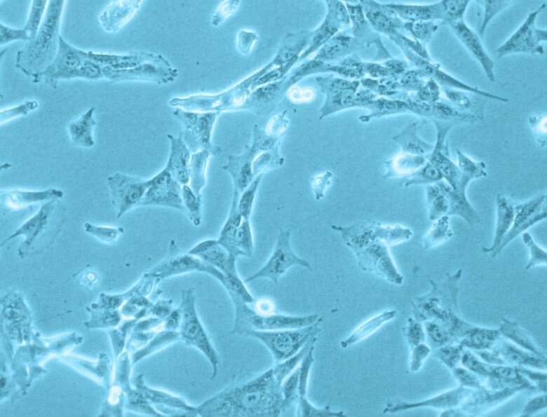
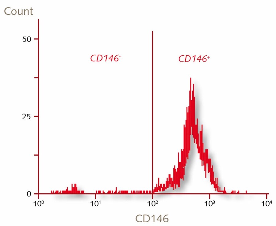
Figure 2. Characterization of human placenta derived vascular pericytes. Human pericytes from placenta cell culture in phase contrast. Flow cytometric analysis of PromoCell’s human placental pericytes are >98% of the cells are CD146+.
Materials
如要继续阅读,请登录或创建帐户。
暂无帐户?