Atto Dyes for Superior Fluorescent Imaging
Activated fluorescent dyes are routinely used to tag proteins, nucleic acids, and other biomolecules for use in life science applications including fluorescence microscopy, flow cytometry, fluorescence in situ hybridization (FISH), receptor binding assays, and enzyme assays. The Atto dyes are a series of fluorescent dyes that meet the critical needs of modern fluorescent technologies:
- Stability - Atto 655 and Atto 647N are photostable and highly resistant to ozone degradation, making them ideal for microarray applications. See the related article "Analyzing Properties of Fluorescent Dyes used for Labeling DNA in Microarray Experiments".
- Long Signal Lifetimes - Signal decay times of 0.6–4.1 nanoseconds allow timegate studies to reduce autofluorescence background and scattering.
- Reduced Background - Several Atto dyes have excitation wavelengths greater than 600 nm, reducing background fluorescence from samples, Rayleigh and Raman scattering.
- Selection - Atto dyes have strong fluorescent signals that cover visible and near-IR emission wavelengths.
Atto Dye's Long Signal Lifetimes
Atto dyes exhibit longer fluorescence signal lifetimes (0.6–4.1 ns) in aqueous solution than either carbocyanine dyes or most of the autofluorescence inherent in cells and biomolecules. The signal from Atto dyes can be measured using pulsed laser excitation with a time-gated detection system to reduce interference from fluorophores with shorter lifetimes, background autofluorescence, and Rayleigh and Raman light scattering, improving overall sensitivity.
Long Excitation Wavelengths Reduces Background
Diode laser excitation at 635 nm and redabsorbing fluorescent dyes were shown to reduce autofluorescence of biological samples sufficiently so that individual antigen and antibody molecules could be detected in human serum samples.1,2 Excitation in the red spectral region also reduces cell damage when working with live cells.3
Many of Atto dyes (Atto 590 and above) can be excited using wavelengths greater than 600 nm. Using long-wavelength activated Atto dyes in conjunction with the appropriate excitation wavelength reduces autofluorescence due to sample, solvent, glass, or polymer support, and improves overall sensitivity in biological analysis and imaging techniques. The background fluorescence due to Rayleigh and Raman scattering are also dramatically reduced by use of longer wavelength excitation.
Emission Wavelength Range from 479 to 764 nm for Fluorescent Multiplex Detection
Atto dyes have strong fluorescent signals with most having molar absorptivity values >100,000 and low excitation/emission overlap, making Atto dyes ideal for multiplex techniques using visible and near-IR emission wavelengths.
With excitation signal maxima ranging from 390 to 740 nm and good Stokes shift separation, there are Atto dyes suitable for use with any common excitation light source.
Fluorescent Multiplex Detection using Atto Dyes
Atto dyes can be used to conjugate probes and biomolecules for multiplex applications. Selection of two Atto dyes with separated emission signals supports multiple excitation and measurement results from a single experiment.
Fluorescent Multiplex Detection using Antibody Atto Dye Conjugates

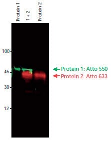
Immunoblot detection of Protein 1 and Protein 2 using two primary antibodies and two anti-IgG-Atto dye conjugates. Imaging was done sequentially using a FLA-3000 Fuji® laser scanner, first at an excitation wavelength of 532 nm with a 580 nm emission filter, then at an excitation wavelength of 633 nm with a 675 nm emission filter. The image overlay was done using a software tool.
Alternatives to Common Fluorophores
With the extensive selection of Atto dyes available, any common excitation light source can be used, and Atto dyes can replace other fluorescent dyes commonly used in life science.
Reactive Atto Dyes and Conjugates
Atto dyes produce intense fluorescent signals due to strong absorbance and high quantum yields. Dyes are available in the following formats:
- Free acid dyes for all routine staining applications
- NHS-esters for use in common conjugation protocols
- Maleimides for use in coupling to thiol-containing groups such as cysteine residues and thiol (−SH) tags added during automated synthesis
- Conjugated to biotin, streptavidin, and antibodies
Atto 655, Atto 680, and Atto 700 are quenched by guanosine, tryptophan and related compounds through direct contact between the dye and the quenching agent and using an electron transfer process. Fluorescent quenching of dyes by tryptophan residues in proteins has been used to differentiate unbound (nonfluorescent) protein from protein-antibody (fluorescent) interactions.1
Convenient Atto Dye Conjugates
An extensive selection of Atto Dye conjugates and kits are available, including:
- Protein Labeling Kits
- Atto 488 is a superior alternative to fluorescein and Alexa Fluor 488, producing conjugates with more photostability and brighter fluorescence.
- Atto 550 is an alternative to rhodamine dyes, Cy3, and Alexa Fluor 550, offering more intense brightness and increased photostability.
- Atto 594 is an alternative to Alexa Fluor 594 and Texas Red.
- Atto 647N is an extraordinary highly fluorescent dye, and Atto 655 are alternatives to Cy5 and Alexa Fluor 647.
- Atto 633 is an alternative to Alexa Fluor 633.
- Lectins for carbohydrate binding studies.
- Primary and Secondary Antibodies for direct and indirect ELISA, Immunoblotting, Immunohistochemistry, and other protein identification applications.
- Biotin and Streptavidin for avidin / streptavidin / biotin conjugation in applications including ELISA, immunohistochemistry, in situ hybridization, and flow cytometry.
- NTA Nickel conjugates for direct detection of polyhistidine-tagged recombinant proteins.

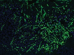
Fluorescent microscopy of human skin tissue section (paraffin fixation) with fungal infection. The target carbohydrate chitotriose of the pathogenic fungi are specifically bound to lectin from Phytolacca americana Atto 488 conjugate (green). The nuclei are counterstained with DAPI (blue). Image by J. Zbären, Inselspital, Bern.

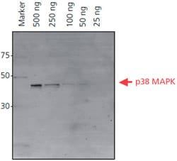
His-tagged p38 MAPK protein (500 ng – 25 ng) was separated on a 4-20% Tris-glycine SDS-PAGE gel. After fixing and washing, the gel was incubated with Ni-NTA-Atto 647N (1:1000) in the dark. The gel was washed and then imaged using a FLA-3000 Fuji® laser scanner with 633 nm excitation and a 675 nm emission filter for Ni-NTA-Atto 647N (λex 647 nm, λem 669 nm). The 50 ng band of His-tagged p38-MAPK is observed using fluorescence imaging.
Atto Dye-labeled Phospholipids
Phospholipids are the major building blocks of biological membranes. The investigation of biological membranes (e.g., intracellular membranes of live cells, plasma membranes) has become a major area of interest. We offer a series of fluorescent-labeled phospholipids. The optical properties of the selected series of dyes allow application with all commonly used excitation and emission filter settings. We offer a variety of phospholipids based on glycerol carrying one or two fatty acids (lipophilic groups) and a phosphate monoester residue (hydrophilic group) such as 1,2-dipalmitoyl-sn-glycero-3-phosphoethanolamine (DPPE), 1,2-dioleoylsn-glycero-3-phosphoethanolamine (DOPE), 1-palmitoyl-2-hydroxy-snglycero-3-phosphoethanolamine (PPE), and 1,2-dimyristoyl-sn-glycero- 3-phosphoethanolamine (DMPE). The fluorophores are covalently linked at the hydrophilic head group of the phospholipids.
Materials
References
如要继续阅读,请登录或创建帐户。
暂无帐户?