Merck
CN
主页癌症研究Prestige Antibodies®在乳腺癌研究中的应用

Prestige Antibodies®在乳腺癌研究中的应用

prestige antibodies-标志
prestige antibodies

人类蛋白质图谱

proteinatlas.org

人类蛋白质图谱表征人类蛋白质组

在克努特和爱丽丝瓦伦贝里基金会(Knut and Alice Wallenberg Foundation)的资助下,由Mathias Uhlén教授领导的瑞典研究团队于2003年发起了人类蛋白质图谱(Human Protein Atlas,HPA)项目。1,2这个独一无二的世界领先级项目利用抗体对人类蛋白质组进行了全面系统的探索。

HPA项目旨在绘制完整人类蛋白质组的表达图谱。为实现该目标,研究人员针对所有蛋白质编码人类基因开发了高度特异性的多克隆抗体,并利用组织阵列技术为大量组织和细胞建立了蛋白质谱图。所采用的应用包括免疫组化(IHC)、Western blot(WB)分析、蛋白质芯片检测和免疫荧光共聚焦显微镜(ICC-IF)。

人类蛋白质图谱,2014年11月

2014年11月发布的人类蛋白质图谱第13版公布了完整人类蛋白质组的组织图谱。大量数据被分为四个独立分册:组织图谱(Tissue Atlas)、癌症图谱(Cancer Atlas)、亚细胞图谱(Subcell Atlas)和细胞系图谱(Cell Line Atlas)。组织图谱中所有蛋白质的表达谱是基于针对大量人类组织的IHC分析, 该图谱现已囊括与每种基因的RNA测序数据相关的蛋白质表达数据。癌症图谱研究了多种癌症中的差异表达基因,而亚细胞图谱提供了通过共聚焦显微镜获得的亚细胞定位。细胞系图谱包含了关于常见细胞系中蛋白质表达的其他信息,该图谱已成为一款备受欢迎的研究工具。

该项目对来自48种不同正常人类组织、20种不同类型癌症和44个不同人类细胞系的样品进行了组织芯片检测。48种正常组织的样品均重复检测三次,并代表82种不同的细胞类型。所有正常组织图像均对表达水平进行了病理学注释并在正常组织图谱中展示,其提供了关于人类基因在mRNA和蛋白质水平上的表达谱的信息。mRNA表达数据来自于27种主要正常组织类型的RNA深度测序(RNA-Seq)数据。

癌症图谱包含基于大量人类癌症标本中蛋白质表达模式的基因表达数据。利用来自216个不同癌症样本的20种最常见人类癌症类型,针对所有包含的基因进行分析。所有癌症组织图片均经过病理学家手动注释,与正常组织图谱一样,蛋白质数据包括对应于16.621个基因的蛋白质表达水平,因为它们均具有可用抗体。

在乳腺组织样本和细胞中进行验证

在正常组织图谱中,每种抗体具有来自三名不同个体的正常乳腺样本的IHC图片。此外,癌症图谱为每种抗体提供了来自多达12名患者的乳腺肿瘤样本(重复2次)的IHC图片,细胞系图谱也为大部分抗体提供了来自MCF-7和SK-BR-3乳腺细胞的图片。

参考文献

1.
Uhlen M, Oksvold P, Fagerberg L, Lundberg E, Jonasson K, Forsberg M, Zwahlen M, Kampf C, Wester K, Hober S, et al. 2010. Towards a knowledge-based Human Protein Atlas. Nat Biotechnol. 28(12):1248-1250. https://doi.org/10.1038/nbt1210-1248
2.
Uhlen M, Ponten F, Lindskog C. 2015. Charting the human proteome: Understanding disease using a tissue-based atlas. Science. 347(6227):1274-1274. https://doi.org/10.1126/science.347.6227.1274-c

Prestige Antibodies® 由Atlas Antibodies提供技术支持

Prestige Antibodies – HPA的基础

Prestige Polyclonals的高度特异性以及与其他蛋白质的低交叉反应性主要基于抗原序列的全面筛选、重组抗原的亲和纯化、多种实验方法验证和严格的审批流程。

显色

Prestige Antibodies的抗原是长度约为50-150个氨基酸的重组人蛋白表位识别标签(PrESTs)。通过使用专门软件对PrESTs进行设计,使这些蛋白质片段含有与天然蛋白质相同的独特表位,并适合激发高特异性抗体的生成。通过人类全基因组扫描,确保作为抗原的PrESTs与其它人类蛋白质的同源性最低。

审批

Prestige Antibodies的审批需综合考量IHC、WB或ICC-IF的实验结果、RNA测序结果以及通过生物信息学预测和参考文献获得的信息。由于文献通常无法判定结论,因此,HPA项目的一个重要目标就是针对同一个蛋白质靶标生产一对无重叠表位的抗体,从而利用这两个抗体相互验证。

Prestige Antibodies产品目录

如今,Prestige Polyclonals的抗体数量已经超过17000个,并且每年新增2,000个抗体。

抗体基于人类蛋白质图谱(HPA)项目开发和表征,以Prestige Antibodies和Atlas Antibodies品牌供给科研人员。


单克隆抗体开发

Prestige Antibodies还包括一定数量的小鼠单克隆抗体。单克隆抗体产品目录每年随新产品的推出而不断扩增。

独特的性质

经过特殊处理的克隆仅能识别独特的无重叠表位和/或同种型。利用与多克隆抗体同样严格的PrEST生产工艺和鉴定流程,使单克隆抗体在获批应用中表现出优异的性能,同时具有明确的特异性、有保证的连续性和稳定的供应。一般来说,这些单克隆抗体可进行高倍数稀释并有助于实现更标准的检测流程。

克隆选择

在对选定杂交瘤进行亚克隆和扩增之前,对大量ELISA阳性细胞上清液进行功能鉴定,为每个应用选择最佳克隆。

表位定位

使用合成重叠肽微珠阵列法对克隆进行表位定位,选出仅含无重叠表位的克隆。

表位定位

使用合成重叠肽微珠阵列法对克隆进行表位定位,选出仅含无重叠表位的克隆。

杂交瘤细胞培养

Atlas Antibodies在放大生产阶段使用体外培养方法,因此无需使用小鼠制备腹水。

抗体鉴定

Prestige单克隆抗体的鉴定从广泛的文献检索开始,选出最相关和最具临床意义的组织用于IHC鉴定。通常,IHC应用数据为每个抗体提供一种以上的组织类型。除阳性染色组织外,还会提供阴性对照组织染色以及相关的临床肿瘤组织染色。

Western blot(WB)鉴定包括来自内源性人类细胞或组织蛋白裂解物或重组全长人类蛋白裂解物(可选)的结果。

因此,每个单克隆抗体均附带其特定靶点的最相关的鉴定数据。

所有Prestige Polyclonals的产品编号以“HPA”开头,单克隆抗体以“AMAB”开头。

临床标记物(ESR1、HER2、Ki67、PGR)

成熟的临床乳腺癌标记物

1.
Pereira CBL, Leal MF, de Souza CRT, Montenegro RC, Rey JA, Carvalho AA, Assumpção PP, Khayat AS, Pinto GR, Demachki S, et al. Prognostic and Predictive Significance of MYC and KRAS Alterations in Breast Cancer from Women Treated with Neoadjuvant Chemotherapy. PLoS ONE. 8(3):e60576. https://doi.org/10.1371/journal.pone.0060576
2.
Camilleri M, Carlson P, Zinsmeister AR, McKinzie S, Busciglio I, Burton D, Zucchelli M, D'Amato M. 2010. Neuropeptide S Receptor Induces Neuropeptide Expression and Associates With Intermediate Phenotypes of Functional Gastrointestinal Disorders. Gastroenterology. 138(1):98-107.e4. https://doi.org/10.1053/j.gastro.2009.08.051
3.
Roca H, Craig MJ, Ying C, Varsos ZS, Czarnieski P, Alva AS, Hernandez J, Fuller D, Daignault S, Healy PN, et al. 2011. IL-4 induces proliferation in prostate cancer PC3?cells under nutrient-depletion stress through the activation of the JNK-pathway and survivin upregulation. J. Cell. Biochem..n/a-n/a. https://doi.org/10.1002/jcb.24025
HER2/ERBB2

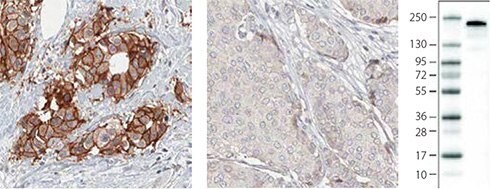
使用HER2抗体(AMAb90627)对人类乳腺肿瘤进行免疫组化染色,结果显示HER2阳性导管癌肿瘤细胞呈强细胞膜阳性(结合中度的细胞质阳性),而HER2阴性导管癌肿瘤细胞无细胞膜阳性。通过Western Blot分析,在乳腺癌细胞SK-BR-3中检测到HER2。

孕激素受体
孕激素受体

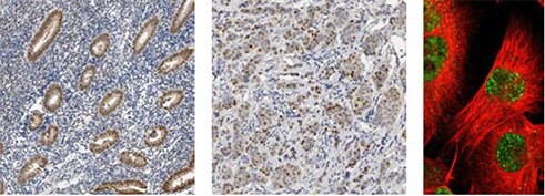
使用PGR抗体(HPA004751)对正常人类宫体(子宫)组织进行IHC染色,结果显示腺细胞呈强细胞核阳性。在提供的乳腺癌样品中,肿瘤细胞染色结果也显示细胞核阳性。ICC-IF结果显示U-251MG细胞核被染色。

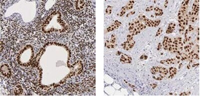
雌激素受体
雌激素受体

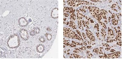
使用ESR1抗体(HPA000449)进行IHC染色,结果显示人乳腺组织中的腺细胞和乳腺癌样品中的肿瘤细胞均呈现明显的细胞核阳性。
雌激素受体-2

使用ESR1抗体(HPA000450)进行IHC染色,结果显示人子宫体组织中的腺细胞和基质细胞以及乳腺癌样品中的肿瘤细胞均呈强细胞核阳性。

Ki67

Ki67

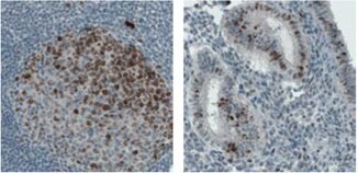
使用MKI67抗体(HPA000451)进行IHC染色,结果显示人淋巴结反应中心的部分细胞呈强细胞核阳性。在乳腺癌中,肿瘤细胞的ICC-IF染色结果也显示细胞核阳性,人U-2OS细胞染色显示核仁阳性。

ki67-2

使用MKI67抗体(HPA001164)对人扁桃体组织进行IHC染色,结果显示反应中心细胞的细胞核被染色。在乳腺癌的肿瘤细胞中,染色主要发生于细胞核,而U-2OS细胞的ICC-IF染色结果显示核仁呈强阳性。
ki67-3

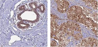
使用单克隆MKI67抗体(AMAb90870)对人结肠淋巴结进行IHC染色,结果显示反应中心细胞呈强细胞核和核仁免疫反应性。子宫组织染色显示部分腺细胞呈细胞核阳性。

乳腺癌研究使用的抗体

本节所述抗体是根据参考文献或与相应靶蛋白的乳腺癌相关性进行选择的。

BRCA1

brca1-1

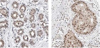
使用BRCA1抗体(HPA034966)进行IHC染色,结果显示正常人乳腺组织中的腺细胞和乳腺癌样品中的肿瘤细胞呈阳性。
brca1-2

使用BRCA2抗体(HPA026815)对正常人乳腺组织进行IHC染色,结果显示腺细胞呈阳性。在乳腺癌样品中,肿瘤细胞呈细胞核染色阳性。

ACAT1
acat1

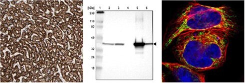
使用ACAT1抗体(HPA004428)对人肝脏组织进行免疫组化染色,结果显示肝细胞呈强细胞质阳性。通过Western Blot分析,在人类细胞RT-4和U251-MG以及肝脏和扁桃体组织裂解物中检测到ACAT1。人类细胞A-431的ICC-IF染色结果显示线粒体呈阳性。

CD44
cd44

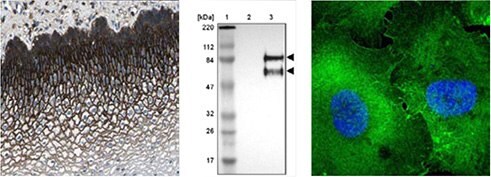
使用CD44抗体(HPA005785)对人食道组织进行免疫组化染色,结果显示鳞状上皮细胞呈强细胞质和细胞膜阳性。通过Western Blot分析,在人类细胞U-251MG中检测到CD44。人类细胞U-251MG的ICC-IF染色结果显示细胞膜呈阳性。

* 对人类和啮齿动物样本均进行WB分析

参考文献

1.
Sanchez-Alvarez R, Martinez-Outschoorn UE, Lin Z, Lamb R, Hulit J, Howell A, Sotgia F, Rubin E, Lisanti MP. 2013. Ethanol exposure induces the cancer-associated fibroblast phenotype and lethal tumor metabolism. Cell Cycle. 12(2):289-301. https://doi.org/10.4161/cc.23109
2.
Martinez-Outschoorn UE, Lin Z, Whitaker-Menezes D, Howell A, Lisanti MP, Sotgia F. 2012. Ketone bodies and two-compartment tumor metabolism: Stromal ketone production fuels mitochondrial biogenesis in epithelial cancer cells. Cell Cycle. 11(21):3956-3963. https://doi.org/10.4161/cc.22136
3.
Martinez-Outschoorn UE, Lin Z, Whitaker-Menezes D, Howell A, Sotgia F, Lisanti MP. 2012. Ketone body utilization drives tumor growth and metastasis. Cell Cycle. 11(21):3964-3971. https://doi.org/10.4161/cc.22137
4.
Chang HT, Olson L, Schwartz KA. 2013. Ketolytic and glycolytic enzymatic expression profiles in malignant gliomas: implication for ketogenic diet therapy. Nutrition & Metabolism. 10(1):47. https://doi.org/10.1186/1743-7075-10-47
5.
Hrstka R, Brychtova V, Fabian P, Vojtesek B, Svoboda M. 2013. AGR2 Predicts Tamoxifen Resistance in Postmenopausal Breast Cancer Patients. Disease Markers. 35207-212. https://doi.org/10.1155/2013/761537
6.
O´Leary PC, Penny SA, Dolan RT, Kelly CM, Madden SF, Rexhepaj E, Brennan DJ, McCann AH, Pontén F, Uhlén M, et al. 2013. Systematic antibody generation and validation via tissue microarray technology leading to identification of a novel protein prognostic panel in breast cancer. BMC Cancer. 13(1): https://doi.org/10.1186/1471-2407-13-175
7.
de Boniface J, Mao Y, Schmidt-Mende J, Kiessling R, Poschke I. 2012. Expression patterns of the immunomodulatory enzyme arginase 1 in blood, lymph nodes and tumor tissue of early-stage breast cancer patients. OncoImmunology. 1(8):1305-1312. https://doi.org/10.4161/onci.21678
8.
Lucki NC, Li D, Bandyopadhyay S, Wang E, Merrill AH, Sewer MB. 2012. Acid Ceramidase (ASAH1) Represses Steroidogenic Factor 1-Dependent Gene Transcription in H295R Human Adrenocortical Cells by Binding to the Receptor. Molecular and Cellular Biology. 32(21):4419-4431. https://doi.org/10.1128/mcb.00378-12
9.
Liang Y, Wu H, Lei R, Chong RA, Wei Y, Lu X, Tagkopoulos I, Kung S, Yang Q, Hu G, et al. 2012. Transcriptional Network Analysis Identifies BACH1 as a Master Regulator of Breast Cancer Bone Metastasis. J. Biol. Chem.. 287(40):33533-33544. https://doi.org/10.1074/jbc.m112.392332
10.
Scheper M, Almubarak H, Jones A, Chaisuparat R, Zhang M, Meiller T. 2011. Zoledronic acid directly suppresses cell proliferation and induces apoptosis in highly tumorigenic prostate and breast cancers. J Carcinog. 10(1):2. https://doi.org/10.4103/1477-3163.75723
11.
Brunquell C, Biliran H, Jennings S, Ireland SK, Chen R, Ruoslahti E. 2012. TLE1 Is an Anoikis Regulator and Is Downregulated by Bit1 in Breast Cancer Cells. Molecular Cancer Research. 10(11):1482-1495. https://doi.org/10.1158/1541-7786.mcr-12-0144
12.
Karmali PP, Brunquell C, Tram H, Ireland SK, Ruoslahti E, Biliran H. Metastasis of Tumor Cells Is Enhanced by Downregulation of Bit1. PLoS ONE. 6(8):e23840. https://doi.org/10.1371/journal.pone.0023840
13.
Vermeulen JF, van Brussel AS, van der Groep P, Morsink FH, Bult P, van der Wall E, van Diest PJ. 2012. Immunophenotyping invasive breast cancer: paving the road for molecular imaging. BMC Cancer. 12(1): https://doi.org/10.1186/1471-2407-12-240
14.
Davidson B, Stavnes HT, Holth A, Chen X, Yang Y, Shih I, Wang T. 2011. Gene expression signatures differentiate ovarian/peritoneal serous carcinoma from breast carcinoma in effusions. 15(3):535-544. https://doi.org/10.1111/j.1582-4934.2010.01019.x
15.
Vermeulen JF, Kornegoor R, van der Wall E, van der Groep P, van Diest PJ. Differential Expression of Growth Factor Receptors and Membrane-Bound Tumor Markers for Imaging in Male and Female Breast Cancer. PLoS ONE. 8(1):e53353. https://doi.org/10.1371/journal.pone.0053353
16.
Tafreshi NK, Bui MM, Bishop K, Lloyd MC, Enkemann SA, Lopez AS, Abrahams D, Carter BW, Vagner J, Grobmyer SR, et al. 2012. Noninvasive Detection of Breast Cancer Lymph Node Metastasis Using Carbonic Anhydrases IX and XII Targeted Imaging Probes. Clinical Cancer Research. 18(1):207-219. https://doi.org/10.1158/1078-0432.ccr-11-0238
17.
Vazquez-Martin A, Oliveras-Ferraros C, Cufí S, Del Barco S, Martin-Castillo B, Menendez JA. 2010. Metformin regulates breast cancer stem cello ntogeny by transcriptional regulation of the epithelial-mesenchymal transition (EMT) status. Cell Cycle. 9(18):3831-3838. https://doi.org/10.4161/cc.9.18.13131
18.
Baccelli I, Schneeweiss A, Riethdorf S, Stenzinger A, Schillert A, Vogel V, Klein C, Saini M, Bäuerle T, Wallwiener M, et al. 2013. Identification of a population of blood circulating tumor cells from breast cancer patients that initiates metastasis in a xenograft assay. Nat Biotechnol. 31(6):539-544. https://doi.org/10.1038/nbt.2576
19.
Petit V, Massonnet G, Maciorowski Z, Touhami J, Thuleau A, Némati F, Laval J, Château-Joubert S, Servely J, Vallerand D, et al. 2013. Optimization of tumor xenograft dissociation for the profiling of cell surface markers and nutrient transporters. Lab Invest. 93(5):611-621. https://doi.org/10.1038/labinvest.2013.44
20.
Twarock S, Röck K, Sarbia M, Weber A, Jänicke R, Fischer J. Synthesis of hyaluronan in oesophageal cancer cells is uncoupled from the prostaglandin-cAMP pathway. 157(2):234-243. https://doi.org/10.1111/j.1476-5381.2009.00138.x
21.
Asplund A, Gry Björklund M, Sundquist C, Strömberg S, Edlund K, Östman A, Nilsson P, Pontén F, Lundeberg J. Expression profiling of microdissected cell populations selected from basal cells in normal epidermis and basal cell carcinoma. 158(3):527-538. https://doi.org/10.1111/j.1365-2133.2007.08418.x
22.
Teleki I, Krenacs T, Szasz MA, Kulka J, Wichmann B, Leo C, Papassotiropoulos B, Riemenschnitter C, Moch H, Varga Z. 2013. The potential prognostic value of connexin 26 and 46 expression in neoadjuvant-treated breast cancer. BMC Cancer. 13(1): https://doi.org/10.1186/1471-2407-13-50
23.
Kiflemariam S, Andersson S, Asplund A, Pontén F, Sjöblom T. Scalable In Situ Hybridization on Tissue Arrays for Validation of Novel Cancer and Tissue-Specific Biomarkers. PLoS ONE. 7(3):e32927. https://doi.org/10.1371/journal.pone.0032927
24.
Nodin B, Fridberg M, Uhlén M, Jirström K. 2012. Discovery of dachshund 2 protein as a novel biomarker of poor prognosis in epithelial ovarian cancer. Journal of Ovarian Research. 5(1):6. https://doi.org/10.1186/1757-2215-5-6
25.
Sircoulomb F, Nicolas N, Ferrari A, Finetti P, Bekhouche I, Rousselet E, Lonigro A, Adélaïde J, Baudelet E, Esteyriès S, et al. 2011. ZNF703 gene amplification at 8p12 specifies luminal B breast cancer. EMBO Mol Med. 3(3):153-166. https://doi.org/10.1002/emmm.201100121
26.
Cawthorn TR, Moreno JC, Dharsee M, Tran-Thanh D, Ackloo S, Zhu PH, Sardana G, Chen J, Kupchak P, Jacks LM, et al. Proteomic Analyses Reveal High Expression of Decorin and Endoplasmin (HSP90B1) Are Associated with Breast Cancer Metastasis and Decreased Survival. PLoS ONE. 7(2):e30992. https://doi.org/10.1371/journal.pone.0030992
27.
Henke A, Grace OC, Ashley GR, Stewart GD, Riddick ACP, Yeun H, O?Donnell M, Anderson RA, Thomson AA. Stromal Expression of Decorin, Semaphorin6D, SPARC, Sprouty1 and Tsukushi in Developing Prostate and Decreased Levels of Decorin in Prostate Cancer. PLoS ONE. 7(8):e42516. https://doi.org/10.1371/journal.pone.0042516
28.
Hudson EP, Uhlen M, Rockberg J. 2012. Multiplex epitope mapping using bacterial surface display reveals both linear and conformational epitopes. Sci Rep. 2(1): https://doi.org/10.1038/srep00706
29.
Arabi A, Ullah K, Branca RM, Johansson J, Bandarra D, Haneklaus M, Fu J, Ariës I, Nilsson P, Den Boer ML, et al. 2012. Proteomic screen reveals Fbw7 as a modulator of the NF-?B pathway. Nat Commun. 3(1): https://doi.org/10.1038/ncomms1975
30.
Ito A, Mimae T, Yamamoto Y, Hagiyama M, Nakanishi J, Ito M, Hosokawa Y, Okada M, Murakami Y, Kondo T. 2012. Novel application for pseudopodia proteomics using excimer laser ablation and two-dimensional difference gel electrophoresis. Lab Invest. 92(9):1374-1385. https://doi.org/10.1038/labinvest.2012.98
31.
Holland DG, Burleigh A, Git A, Goldgraben MA, Perez?Mancera PA, Chin S, Hurtado A, Bruna A, Ali HR, Greenwood W, et al. 2011. ZNF703 is a common Luminal B breast cancer oncogene that differentially regulates luminal and basal progenitors in human mammary epithelium. EMBO Mol Med. 3(3):167-180. https://doi.org/10.1002/emmm.201100122
32.
Mulder J, Björling E, Jonasson K, Wernérus H, Hober S, Hökfelt T, Uhlén M. 2009. Tissue Profiling of the Mammalian Central Nervous System Using Human Antibody-based Proteomics. Mol Cell Proteomics. 8(7):1612-1622. https://doi.org/10.1074/mcp.m800539-mcp200
33.
Su D, Fu X, Fan S, Wu X, Wang X, Fu L, Dong X, Ni JJ, Fu L, Zhu Z, et al. 2012. Role of ERRF, a Novel ER-Related Nuclear Factor, in the Growth Control of ER-Positive Human Breast Cancer Cells. The American Journal of Pathology. 180(3):1189-1201. https://doi.org/10.1016/j.ajpath.2011.11.025
34.
Shubbar E, Helou K, Kovács A, Nemes S, Hajizadeh S, Enerbäck C, Einbeigi Z. 2013. High levels of ?-glutamyl hydrolase (GGH) are associated with poor prognosis and unfavorable clinical outcomes in invasive breast cancer. BMC Cancer. 13(1): https://doi.org/10.1186/1471-2407-13-47
35.
Liao YC, Ruan JW, Lua I, Li MH, Chen WL, Wang JRY, Kao RH, Chen JH. 2012. Overexpressed hPTTG1 promotes breast cancer cell invasion and metastasis by regulating GEF-H1/RhoA signalling. Oncogene. 31(25):3086-3097. https://doi.org/10.1038/onc.2011.476
36.
Cheng IK, Tsang BC, Lai KP, Ching AK, Chan AW, To K, Lai PB, Wong N. 2012. GEF-H1 over-expression in hepatocellular carcinoma promotes cell motility via activation of RhoA signalling. J. Pathol.. 228(4):575-585. https://doi.org/10.1002/path.4084
37.
Zibert JR, Wallbrecht K, Schön M, Mir LM, Jacobsen GK, Trochon-Joseph V, Bouquet C, Villadsen LS, Cadossi R, Skov L, et al. 2011. Halting angiogenesis by non-viral somatic gene therapy alleviates psoriasis and murine psoriasiform skin lesions. J. Clin. Invest.. 121(1):410-421. https://doi.org/10.1172/jci41295
38.
Smyth LG, O'Hurley G, O'Grady A, Fitzpatrick JM, Kay E, Watson RWG. 2010. Carbonic anhydrase IX expression in prostate cancer. Prostate Cancer Prostatic Dis. 13(2):178-181. https://doi.org/10.1038/pcan.2009.58
39.
Paatero I, Jokilammi A, Heikkinen PT, Iljin K, Kallioniemi O, Jones FE, Jaakkola PM, Elenius K. 2012. Interaction with ErbB4 Promotes Hypoxia-inducible Factor-1? Signaling. J. Biol. Chem.. 287(13):9659-9671. https://doi.org/10.1074/jbc.m111.299537
40.
Zbytek B, Peacock DL, Seagroves TN, Slominski A. 2013. Putative role of HIF transcriptional activity in melanocytes and melanoma biology. Dermato-Endocrinology. 5(2):239-251. https://doi.org/10.4161/derm.22678
41.
Hu Z, Huang G, Sadanandam A, Gu S, Lenburg ME, Pai M, Bayani N, Blakely EA, Gray JW, Mao J. 2010. The expression level of HJURP has an independent prognostic impact and predicts the sensitivity to radiotherapy in breast cancer. Breast Cancer Res. 12(2): https://doi.org/10.1186/bcr2487
42.
Shuaib M, Ouararhni K, Dimitrov S, Hamiche A. 2010. HJURP binds CENP-A via a highly conserved N-terminal domain and mediates its deposition at centromeres. Proceedings of the National Academy of Sciences. 107(4):1349-1354. https://doi.org/10.1073/pnas.0913709107
43.
de Tayrac M, Saikali S, Aubry M, Bellaud P, Boniface R, Quillien V, Mosser J. Prognostic Significance of EDN/RB, HJURP, p60/CAF-1 and PDLI4, Four New Markers in High-Grade Gliomas. PLoS ONE. 8(9):e73332. https://doi.org/10.1371/journal.pone.0073332
44.
Bjarnadottir O, Romero Q, Bendahl P, Jirström K, Rydén L, Loman N, Uhlén M, Johannesson H, Rose C, Grabau D, et al. 2013. Targeting HMG-CoA reductase with statins in a window-of-opportunity breast cancer trial. Breast Cancer Res Treat. 138(2):499-508. https://doi.org/10.1007/s10549-013-2473-6
45.
Jaraj SJ, Augsten M, Häggarth L, Wester K, Pontén F, Östman A, Egevad L. 2011. GAD1 is a biomarker for benign and malignant prostatic tissue. Scandinavian Journal of Urology and Nephrology. 45(1):39-45. https://doi.org/10.3109/00365599.2010.521189
46.
Liu H, Zhang W, Jia Y, Yu Q, Grau GE, Peng L, Ran Y, Yang Z, Deng H, Lou J. 2013. Single-cell clones of liver cancer stem cells have the potential of differentiating into different types of tumor cells. Cell Death Dis. 4(10):e857-e857. https://doi.org/10.1038/cddis.2013.340
47.
Guo L, Chen C, Shi M, Wang F, Chen X, Diao D, Hu M, Yu M, Qian L, Guo N. 2013. Stat3-coordinated Lin-28?let-7?HMGA2 and miR-200?ZEB1 circuits initiate and maintain oncostatin M-driven epithelial?mesenchymal transition. Oncogene. 32(45):5272-5282. https://doi.org/10.1038/onc.2012.573
48.
Possemato R, Marks KM, Shaul YD, Pacold ME, Kim D, Birsoy K, Sethumadhavan S, Woo H, Jang HG, Jha AK, et al. 2011. Functional genomics reveal that the serine synthesis pathway is essential in breast cancer. Nature. 476(7360):346-350. https://doi.org/10.1038/nature10350
49.
Maddocks ODK, Berkers CR, Mason SM, Zheng L, Blyth K, Gottlieb E, Vousden KH. 2013. Serine starvation induces stress and p53-dependent metabolic remodelling in cancer cells. Nature. 493(7433):542-546. https://doi.org/10.1038/nature11743
50.
Nilsson LM, Plym Forshell TZ, Rimpi S, Kreutzer C, Pretsch W, Bornkamm GW, Nilsson JA. Mouse Genetics Suggests Cell-Context Dependency for Myc-Regulated Metabolic Enzymes during Tumorigenesis. PLoS Genet. 8(3):e1002573. https://doi.org/10.1371/journal.pgen.1002573
51.
Salem AF, Whitaker-Menezes D, Howell A, Sotgia F, Lisanti MP. 2012. Mitochondrial biogenesis in epithelial cancer cells promotes breast cancer tumor growth and confers autophagy resistance. Cell Cycle. 11(22):4174-4180. https://doi.org/10.4161/cc.22376
52.
Wagoner MP, Gunsalus KTW, Schoenike B, Richardson AL, Friedl A, Roopra A. The Transcription Factor REST Is Lost in Aggressive Breast Cancer. PLoS Genet. 6(6):e1000979. https://doi.org/10.1371/journal.pgen.1000979
53.
Prada I, Marchaland J, Podini P, Magrassi L, D'Alessandro R, Bezzi P, Meldolesi J. 2011. REST/NRSF governs the expression of dense-core vesicle gliosecretion in astrocytes. 193(3):537-549. https://doi.org/10.1083/jcb.201010126
54.
Jögi A, Brennan DJ, Rydén L, Magnusson K, Fernö M, Stål O, Borgquist S, Uhlen M, Landberg G, Påhlman S, et al. 2009. Nuclear expression of the RNA-binding protein RBM3 is associated with an improved clinical outcome in breast cancer. Mod Pathol. 22(12):1564-1574. https://doi.org/10.1038/modpathol.2009.124
55.
Hjelm B, Brennan DJ, Zendehrokh N, Eberhard J, Nodin B, Gaber A, Pontén F, Johannesson H, Smaragdi K, Frantz C, et al. 2011. High nuclear RBM3 expression is associated with an improved prognosis in colorectal cancer. Prot. Clin. Appl.. 5(11-12):624-635. https://doi.org/10.1002/prca.201100020
56.
Ehlén Å, Brennan DJ, Nodin B, O'Connor DP, Eberhard J, Alvarado-Kristensson M, Jeffrey IB, Manjer J, Brändstedt J, Uhlén M, et al. 2010. Expression of the RNA-binding protein RBM3 is associated with a favourable prognosis and cisplatin sensitivity in epithelial ovarian cancer. Journal of Translational Medicine. 8(1):78. https://doi.org/10.1186/1479-5876-8-78
57.
Jonsson L, Gaber A, Ulmert D, Uhlén M, Bjartell A, Jirström K. 2011. High RBM3 expression in prostate cancer independently predicts a reduced risk of biochemical recurrence and disease progression. Diagn Pathol. 6(1): https://doi.org/10.1186/1746-1596-6-91
58.
Nodin B, Fridberg M, Jonsson L, Bergman J, Uhlén M, Jirström K. 2012. High MCM3 expression is an independent biomarker of poor prognosis and correlates with reduced RBM3 expression in a prospective cohort of malignant melanoma. Diagnostic Pathology. 7(1):82. https://doi.org/10.1186/1746-1596-7-82
59.
Jonsson L, Bergman J, Nodin B, Manjer J, Pontén F, Uhlén M, Jirström K. 2011. Low RBM3 protein expression correlates with tumour progression and poor prognosis in malignant melanoma: An analysis of 215 cases from the Malmö Diet and Cancer Study. Journal of Translational Medicine. 9(1):114. https://doi.org/10.1186/1479-5876-9-114
60.
Ehlén Õ, Nodin B, Rexhepaj E, Brändstedt J, Uhlén M, Alvarado-Kristensson M, Pontén F, Brennan DJ, Jirström K. 2011. RBM3-Regulated Genes Promote DNA Integrity and Affect Clinical Outcome in Epithelial Ovarian Cancer. Translational Oncology. 4(4):212-IN1. https://doi.org/10.1593/tlo.11106
61.
Hjelm B, Brennan DJ, Zendehrokh N, Eberhard J, Nodin B, Gaber A, Pontén F, Johannesson H, Smaragdi K, Frantz C, et al. 2011. High nuclear RBM3 expression is associated with an improved prognosis in colorectal cancer. Prot. Clin. Appl.. 5(11-12):624-635. https://doi.org/10.1002/prca.201100020
62.
Boman K, Segersten U, Ahlgren G, Eberhard J, Uhlén M, Jirström K, Malmström P. 2013. Decreased expression of RNA-binding motif protein 3 correlates with tumour progression and poor prognosis in urothelial bladder cancer. BMC Urol. 13(1): https://doi.org/10.1186/1471-2490-13-17
63.
Nodin B, Fridberg M, Jonsson L, Bergman J, Uhlén M, Jirström K. 2012. High MCM3 expression is an independent biomarker of poor prognosis and correlates with reduced RBM3 expression in a prospective cohort of malignant melanoma. Diagnostic Pathology. 7(1):82. https://doi.org/10.1186/1746-1596-7-82
64.
Telikicherla D, Marimuthu A, Kashyap M, Ramachandra YL, Mohan S, Roa J, Maharudraiah J, Pandey A. 2012. Overexpression of ribosome binding protein 1 (RRBP1) in breast cancer. Clin Proteomics. 9(1):7. https://doi.org/10.1186/1559-0275-9-7
65.
Iwanaga R, Wang C, Micalizzi DS, Harrell JC, Jedlicka P, Sartorius CA, Kabos P, Farabaugh SM, Bradford AP, Ford HL. 2012. Expression of Six1 in luminal breast cancers predicts poor prognosis and promotes increases in tumor initiating cells by activation of extracellular signal-regulated kinase and transforming growth factor-beta signaling pathways. Breast Cancer Res. 14(4): https://doi.org/10.1186/bcr3219
66.
Smith AL, Iwanaga R, Drasin DJ, Micalizzi DS, Vartuli RL, Tan A, Ford HL. 2012. The miR-106b-25 cluster targets Smad7, activates TGF-? signaling, and induces EMT and tumor initiating cell characteristics downstream of Six1 in human breast cancer. Oncogene. 31(50):5162-5171. https://doi.org/10.1038/onc.2012.11
67.
Wan F, Miao X, Quraishi I, Kennedy V, Creek KE, Pirisi L. 2008. Gene expression changes during HPV-mediated carcinogenesis: A comparison between anin vitrocell model and cervical cancer. Int. J. Cancer. 123(1):32-40. https://doi.org/10.1002/ijc.23463
68.
McCoy EL, Iwanaga R, Jedlicka P, Abbey N, Chodosh LA, Heichman KA, Welm AL, Ford HL. 2009. Six1 expands the mouse mammary epithelial stem/progenitor cell pool and induces mammary tumors that undergo epithelial-mesenchymal transition. J. Clin. Invest.. 119(9):2663-2677. https://doi.org/10.1172/jci37691
69.
Micalizzi DS, Christensen KL, Jedlicka P, Coletta RD, Barón AE, Harrell JC, Horwitz KB, Billheimer D, Heichman KA, Welm AL, et al. 2009. The Six1 homeoprotein induces human mammary carcinoma cells to undergo epithelial-mesenchymal transition and metastasis in mice through increasing TGF-? signaling. J. Clin. Invest.. 119(9):2678-2690. https://doi.org/10.1172/jci37815
70.
Farabaugh SM, Micalizzi DS, Jedlicka P, Zhao R, Ford HL. 2012. Eya2 is required to mediate the pro-metastatic functions of Six1 via the induction of TGF-? signaling, epithelial?mesenchymal transition, and cancer stem cell properties. Oncogene. 31(5):552-562. https://doi.org/10.1038/onc.2011.259
71.
Ono H, Imoto I, Kozaki K, Tsuda H, Matsui T, Kurasawa Y, Muramatsu T, Sugihara K, Inazawa J. 2012. SIX1 promotes epithelial?mesenchymal transition in colorectal cancer through ZEB1 activation. Oncogene. 31(47):4923-4934. https://doi.org/10.1038/onc.2011.646
72.
Le Grand F, Grifone R, Mourikis P, Houbron C, Gigaud C, Pujol J, Maillet M, Pagès G, Rudnicki M, Tajbakhsh S, et al. 2012. Six1 regulates stem cell repair potential and self-renewal during skeletal muscle regeneration. 198(5):815-832. https://doi.org/10.1083/jcb.201201050
73.
Sernbo S, Borrebaeck CAK, Uhlén M, Jirström K, Ek S. Nuclear T-STAR Protein Expression Correlates with HER2 Status, Hormone Receptor Negativity and Prolonged Recurrence Free Survival in Primary Breast Cancer and Decreased Cancer Cell Growth In Vitro. PLoS ONE. 8(7):e70596. https://doi.org/10.1371/journal.pone.0070596
74.
Ek S, Andréasson U, Hober S, Kampf C, Pontén F, Uhlén M, Merz H, Borrebaeck CAK. 2006. From Gene Expression Analysis to Tissue Microarrays. Mol Cell Proteomics. 5(6):1072-1081. https://doi.org/10.1074/mcp.m600077-mcp200
75.
Schenke-Layland K, Stock UA, Nsair A, Xie J, Angelis E, Fonseca CG, Larbig R, Mahajan A, Shivkumar K, Fishbein MC, et al. 2009. Cardiomyopathy is associated with structural remodelling of heart valve extracellular matrix. 30(18):2254-2265. https://doi.org/10.1093/eurheartj/ehp267
76.
Ghosh Z, Huang M, Hu S, Wilson KD, Dey D, Wu JC. 2011. Dissecting the Oncogenic and Tumorigenic Potential of Differentiated Human Induced Pluripotent Stem Cells and Human Embryonic Stem Cells. Cancer Research. 71(14):5030-5039. https://doi.org/10.1158/0008-5472.can-10-4402
77.
Edlund K, Lindskog C, Saito A, Berglund A, Pontén F, Göransson-Kultima H, Isaksson A, Jirström K, Planck M, Johansson L, et al. 2012. CD99 is a novel prognostic stromal marker in non-small cell lung cancer. Int. J. Cancer. 131(10):2264-2273. https://doi.org/10.1002/ijc.27518
78.
Pontén F, Jirström K, Uhlen M. 2008. The Human Protein Atlas?a tool for pathology. J. Pathol.. 216(4):387-393. https://doi.org/10.1002/path.2440
79.
Wu C, Hsu C, Chen C, Yu C, Chang K, Tai D, Liu H, Su W, Chang Y, Yu J. 2010. Candidate Serological Biomarkers for Cancer Identified from the Secretomes of 23 Cancer Cell Lines and the Human Protein Atlas. Mol Cell Proteomics. 9(6):1100-1117. https://doi.org/10.1074/mcp.m900398-mcp200
80.
Davidson B, Stavnes HT, Holth A, Chen X, Yang Y, Shih I, Wang T. 2011. Gene expression signatures differentiate ovarian/peritoneal serous carcinoma from breast carcinoma in effusions. 15(3):535-544. https://doi.org/10.1111/j.1582-4934.2010.01019.x
81.
Dzi?giel P, Owczarek T, Plaz?uk E, Gomu?kiewicz A, Majchrzak M, Podhorska-Oko?ów M, Driouch K, Lidereau R, Ugorski M. 2010. Ceramide galactosyltransferase (UGT8) is a molecular marker of breast cancer malignancy and lung metastases. Br J Cancer. 103(4):524-531. https://doi.org/10.1038/sj.bjc.6605750
82.
de Kruijf EM, Sajet A, van Nes JG, Putter H, Smit VT, Eagle RA, Jafferji I, Trowsdale J, Liefers GJ, van de Velde CJ, et al. 2012. NKG2D ligand tumor expression and association with clinical outcome in early breast cancer patients: an observational study. BMC Cancer. 12(1): https://doi.org/10.1186/1471-2407-12-24
83.
Elkabets M, Gifford AM, Scheel C, Nilsson B, Reinhardt F, Bray M, Carpenter AE, Jirström K, Magnusson K, Ebert BL, et al. 2011. Human tumors instigate granulin-expressing hematopoietic cells that promote malignancy by activating stromal fibroblasts in mice. J. Clin. Invest.. 121(2):784-799. https://doi.org/10.1172/jci43757

MammaPrint、Oncotype、EndoPredict和uPA检测中基因产物的抗体

本节介绍了Prestige Antibody产品目录中靶向诊断性MammaPrint、Oncotype、EndoPredict和uPA检测所含基因产物的抗体。MammaPrint是一项基因表达谱测试,基于Agendia 公司推出的Amsterdam 70-基因乳腺癌基因标记检测。该测试可评估乳腺癌向身体其他部位转移的风险。MammaPrint的目标是将患者分为“低风险”和“高风险”类别。Oncotype DX(由Genomic Health开发)是美国临床实践中最常用的基因表达谱分析方法,它能够检测肿瘤中的21个基因并确定复发分数(Recurrence Score)。

BIRC5/Survivin
BIRC5/Survivin

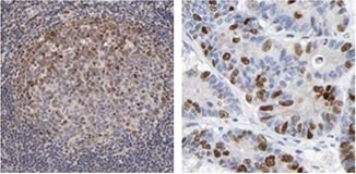
使用BIRC5抗体(HPA002830)进行IHC染色,结果显示人扁桃体组织中的生发中心细胞和结直肠中的肿瘤细胞呈细胞核阳性。

CD68/Macrosialin
CD68/Macrosialin

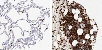
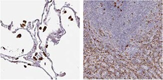
使用CD68抗体(HPA048982)对人肺组织进行IHC染色,结果显示巨噬细胞和造血组织(如脾脏)呈强细胞质阳性。

DTL
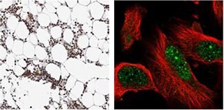
DTL

使用DTL抗体(HPA028016)对人骨髓进行IHC染色,结果显示骨髓造血细胞呈强细胞核阳性。通过ICC-IF染色,检测到U-251 MG细胞的细胞核被染色。

GSTM5
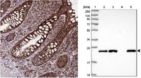
GSTM5

使用GSTM5抗体(HPA048652)进行IHC染色,结果显示人直肠腺细胞呈细胞质阳性;同时,通过WB分析,在RT-4和U-251 MG细胞裂解物以及肝脏组织裂解物中检测到预期大小的条带。

* 对人类和啮齿动物样本均进行WB分析

1.
Gill RM, Gabor TV, Couzens AL, Scheid MP. 2013. The MYC-Associated Protein CDCA7 Is Phosphorylated by AKT To Regulate MYC-Dependent Apoptosis and Transformation. Mol. Cell. Biol.. 33(3):498-513. https://doi.org/10.1128/mcb.00276-12
2.
Shubbar E, Kovács A, Hajizadeh S, Parris TZ, Nemes S, Gunnarsdóttir K, Einbeigi Z, Karlsson P, Helou K. 2013. Elevated cyclin B2 expression in invasive breast carcinoma is associated with unfavorable clinical outcome. BMC Cancer. 13(1): https://doi.org/10.1186/1471-2407-13-1
3.
Karaayvaz M, Pal T, Song B, Zhang C, Georgakopoulos P, Mehmood S, Burke S, Shroyer K, Ju J. 2011. Prognostic Significance of miR-215 in Colon Cancer. Clinical Colorectal Cancer. 10(4):340-347. https://doi.org/10.1016/j.clcc.2011.06.002
4.
Bozóky B, Savchenko A, Csermely P, Korcsmáros T, Dúl Z, Pontén F, Székely L, Klein G. 2013. Novel signatures of cancer-associated fibroblasts. Int. J. Cancer. 133(2):286-293. https://doi.org/10.1002/ijc.28035
5.
Rognum IJ, Haynes RL, Vege ?, Yang M, Rognum TO, Kinney HC. 2009. Interleukin-6 and the serotonergic system of the medulla oblongata in the sudden infant death syndrome. Acta Neuropathol. 118(4):519-530. https://doi.org/10.1007/s00401-009-0535-y

MMP9

mmp9-1

使用MMP9抗体(HPA001238)对人肺组织进行IHC染色,结果显示骨髓组织中的巨噬细胞和骨髓造血细胞呈强细胞核阳性。
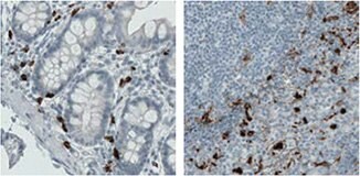
mmp9-2

使用单克隆MMP9抗体染色,结果显示十二指肠(AMAb90805)和人扁桃体组织(AMAb90804)中的部分淋巴细胞显示强细胞质阳性。

LYRIC/MTDH

lyric-mtdh-1

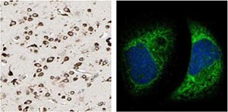
使用MTDH抗体(HPA010932)进行IHC染色,结果显示人大脑皮层组织中的神经元细胞呈强细胞质阳性。对A-431细胞的ICC-IF染色结果显示,内质网被抗体染色。
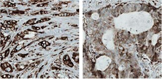
lyric-mtdh-2

使用单克隆MTDH抗体(AMAb90762)进行IHC染色,结果显示乳腺和结直肠癌样品中的肿瘤细胞呈强细胞质反应性。

P5C脱氢酶/ALDH4A1
p5c-脱氢酶-aldh4a1

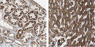
使用ALDH4A1抗体(HPA006401)进行IHC染色,结果显示人肾脏和肝脏组织呈强细胞质阳性并具有颗粒性。

* 对人类和啮齿动物样本均进行WB分析

1.
Pohler E, Mamai O, Hirst J, Zamiri M, Horn H, Nomura T, Irvine AD, Moran B, Wilson NJ, Smith FJD, et al. 2012. Haploinsufficiency for AAGAB causes clinically heterogeneous forms of punctate palmoplantar keratoderma. Nat Genet. 44(11):1272-1276. https://doi.org/10.1038/ng.2444
2.
Roca H, Craig MJ, Ying C, Varsos ZS, Czarnieski P, Alva AS, Hernandez J, Fuller D, Daignault S, Healy PN, et al. 2011. IL-4 induces proliferation in prostate cancer PC3?cells under nutrient-depletion stress through the activation of the JNK-pathway and survivin upregulation. J. Cell. Biochem..n/a-n/a. https://doi.org/10.1002/jcb.24025
3.
Friedman JS, Chang B, Krauth DS, Lopez I, Waseem NH, Hurd RE, Feathers KL, Branham KE, Shaw M, Thomas GE, et al. 2010. Loss of lysophosphatidylcholine acyltransferase 1 leads to photoreceptor degeneration in rd11 mice. Proceedings of the National Academy of Sciences. 107(35):15523-15528. https://doi.org/10.1073/pnas.1002897107
4.
Nohata N, Hanazawa T, Kikkawa N, Mutallip M, Sakurai D, Fujimura L, Kawakami K, Chiyomaru T, Yoshino H, Enokida H, et al. 2011. Tumor suppressive microRNA-375 regulates oncogene AEG-1/MTDH in head and neck squamous cell carcinoma (HNSCC). J Hum Genet. 56(8):595-601. https://doi.org/10.1038/jhg.2011.66
5.
LIU B, WU Y, PENG D. 2013. Astrocyte elevated gene-1 regulates osteosarcoma cell invasion and chemoresistance via endothelin-1/endothelin A receptor signaling. 5(2):505-510. https://doi.org/10.3892/ol.2012.1056
6.
Lorenzen JM, Martino F, Scheffner I, Bröcker V, Leitolf H, Haller H, Gwinner W. Fetuin, Matrix-Gla Protein and Osteopontin in Calcification of Renal Allografts. PLoS ONE. 7(12):e52039. https://doi.org/10.1371/journal.pone.0052039

PITRM1/MP1

pitrm1-mp1

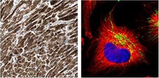
使用PITRM1抗体(HPA006753)进行IHC染色,结果显示人心肌细胞呈强细胞质阳性。人类细胞U-251 MG的ICC-IF染色结果显示线粒体呈阳性。

PRC1
prc1

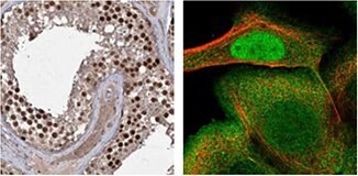
使用PRC1抗体(HPA034521)对人睾丸组织进行IHC染色,结果显示生精小管的细胞呈强细胞核阳性。A-431细胞的ICC-IF染色显示细胞核、细胞膜和微管被染色。

SCOT/OXCT1

scot-oxct1

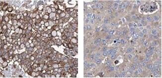
使用OXCT1抗体(HPA012047)对人心肌和肾脏进行IHC染色,结果显示心肌细胞和肾小管细胞分别呈强细胞质阳性。A-431细胞的ICC-IF染色显示线粒体被染色。

* 对人类和啮齿动物样本均进行WB分析
1.
Pereira CBL, Leal MF, de Souza CRT, Montenegro RC, Rey JA, Carvalho AA, Assumpção PP, Khayat AS, Pinto GR, Demachki S, et al. Prognostic and Predictive Significance of MYC and KRAS Alterations in Breast Cancer from Women Treated with Neoadjuvant Chemotherapy. PLoS ONE. 8(3):e60576. https://doi.org/10.1371/journal.pone.0060576
2.
Chang HT, Olson L, Schwartz KA. 2013. Ketolytic and glycolytic enzymatic expression profiles in malignant gliomas: implication for ketogenic diet therapy. Nutrition & Metabolism. 10(1):47. https://doi.org/10.1186/1743-7075-10-47
3.
Zibert JR, Wallbrecht K, Schön M, Mir LM, Jacobsen GK, Trochon-Joseph V, Bouquet C, Villadsen LS, Cadossi R, Skov L, et al. 2011. Halting angiogenesis by non-viral somatic gene therapy alleviates psoriasis and murine psoriasiform skin lesions. J. Clin. Invest.. 121(1):410-421. https://doi.org/10.1172/jci41295

在人类蛋白图谱中鉴定的抗体

在乳腺癌样本中展现出差异的IHC染色模式

抗-klhl26-抗体

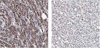
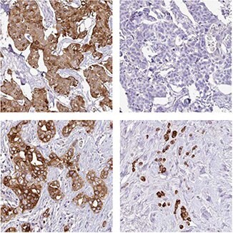
采用抗KLHL26抗体(HPA023074)进行IHC分析,结果显示不同病人的乳腺癌样本具有不同的膜或胞浆染色模式。
抗-acsf2-抗体

ACSF2抗体(HPA024693)表明在不同病人的乳腺癌细胞中具有颗粒蛋白胞浆阳性,并呈强至阴性。
抗-gcm1-抗体

抗GCM1抗体(HPA011343)分析在乳腺癌细胞中呈膜阳性但在正常乳腺组织中为阴性。
抗-agr3-抗体

抗AGR3抗体(HPA053942)表明在11/12例乳腺癌病人中具有强的胞浆阳性而1例病人是完全的阴性。

* 对人类和啮齿动物样本均进行WB分析

1.
Ngan E, Diamond B. 2012. LPP is Required for TGF-Beta Induced Motility and Invasion of Neu/ErbB-2 Expressing Breast Cancer Cells. https://doi.org/10.21236/ada568114
2.
Camilleri M, Carlson P, Zinsmeister AR, McKinzie S, Busciglio I, Burton D, Zucchelli M, D'Amato M. 2010. Neuropeptide S Receptor Induces Neuropeptide Expression and Associates With Intermediate Phenotypes of Functional Gastrointestinal Disorders. Gastroenterology. 138(1):98-107.e4. https://doi.org/10.1053/j.gastro.2009.08.051
3.
Bozóky B, Savchenko A, Csermely P, Korcsmáros T, Dúl Z, Pontén F, Székely L, Klein G. 2013. Novel signatures of cancer-associated fibroblasts. Int. J. Cancer. 133(2):286-293. https://doi.org/10.1002/ijc.28035
4.
Stro?mberg S, Agnarsdo?ttir M, Magnusson K, Rexhepaj E, Bolander Å, Lundberg E, Asplund A, Ryan D, Rafferty M, Gallagher WM, et al. 2009. Selective Expression of Syntaxin-7 Protein in Benign Melanocytes and Malignant Melanoma. J. Proteome Res.. 8(4):1639-1646. https://doi.org/10.1021/pr800745e

乳腺癌

乳腺癌作为第二常见的癌症,是迄今为止女性中最常见的癌症。乳腺癌的发病率正稳步上升,但死亡率并没有相应增加。如果能在早期被检测到,对于生活在发达国家的患者预后相对较好,一般五年生存率约为85%。

乳腺癌及治疗

虽然癌症通常被称为单一疾病,但它确实是一种多维度疾病。多年来,这种理解已经在发生变化,但许多患者并未接受针对其疾病的最佳治疗。对于癌症患者接受更个性化的治疗,仍然需要新的和更好的方法来对患者进行分层。经典的预后因素如肿瘤分期和分级并不足以对患者的预后进行正确的预估。来自癌症生物标志物的其他信息有望对这种预估作出极大的改善,并最终导致更为个性化的治疗,从而避免对患者进行治疗不足和过度治疗。

乳腺癌患者的主要治疗方法是手术,并通常与辅助治疗相结合。然而,辅助治疗与实质性成本相关并有时会伴有严重的副作用,而且医生已经将过度治疗的减少确定为当今乳腺癌治疗中的主要临床需要。因此,将患者分层为不同的预后类别是非常重要的,因为它可以帮助医生为给定患者选择最为合适的治疗方案。

大多数乳腺癌是激素受体反应性的,即表达雌激素受体(ER)或孕酮受体(PR)。带有这些受体表达的肿瘤患者可以接受辅助内分泌治疗,例如他莫昔芬。

乳腺癌也可表达HER2蛋白(人表皮生长因子受体2),表达该蛋白的肿瘤患者可接受曲妥珠单抗的辅助治疗。

辅助治疗也可以包括化学疗法或放射疗法。

RBM3

RNA结合基序蛋白3(RBM3)是一种RNA和DNA结合蛋白,其功能尚未完全阐明。该蛋白质已被证明在亚低温中可作为一种早期事件而实现表达,并也在与细胞应激相关的其他条件下发生表达,例如葡萄糖剥夺和缺氧。在应激条件下,RBM3被认为可通过帮助维持生存所需的蛋白合成来对细胞实现保护1。近期研究还显示RBM3可减弱前列腺癌细胞中的干细胞样特性2

通过人类蛋白质图谱(HPA),RBM3作为一种潜在的肿瘤学生物标志物可通过作为HPA项目 (proteinatlas.org)的一部分在被调查的几种癌症中因为存在差异表达模式而得以鉴定3,4

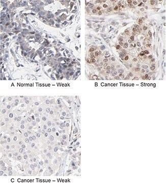
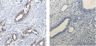
使用抗RBM3抗体HPA003624的IHC分析显示其在正常乳腺组织中的弱表达模式,但在乳腺癌组织中则呈分层模式(图1)。研究人员还进一步研究了其在较大乳腺癌队列中的表达,以及RBM3的表达显示出与延长的生存率相关5

1.
Ehlén Å. 2011. Te Role of RNA-Binding Motif 3 in Epithelial Ovarian Cancer: A Biomarker Discovery Approach .
2.
Zeng Y, Wodzenski D, Gao D, Shiraishi T, Terada N, Li Y, Vander Griend DJ, Luo J, Kong C, Getzenberg RH, et al. 2013. Stress-Response Protein RBM3 Attenuates the Stem-like Properties of Prostate Cancer Cells by Interfering with CD44 Variant Splicing. Cancer Res. 73(13):4123-4133. https://doi.org/10.1158/0008-5472.can-12-1343
3.
Berglund L, Björling E, Oksvold P, Fagerberg L, Asplund A, Al-Khalili Szigyarto C, Persson A, Ottosson J, Wernérus H, Nilsson P, et al. 2008. A Genecentric Human Protein Atlas for Expression Profiles Based on Antibodies. Mol Cell Proteomics. 7(10):2019-2027. https://doi.org/10.1074/mcp.r800013-mcp200
4.
Uhlen M, Oksvold P, Fagerberg L, Lundberg E, Jonasson K, Forsberg M, Zwahlen M, Kampf C, Wester K, Hober S, et al. 2010. Towards a knowledge-based Human Protein Atlas. Nat Biotechnol. 28(12):1248-1250. https://doi.org/10.1038/nbt1210-1248
5.
Jögi A, Brennan DJ, Rydén L, Magnusson K, Fernö M, Stål O, Borgquist S, Uhlen M, Landberg G, Påhlman S, et al. 2009. Nuclear expression of the RNA-binding protein RBM3 is associated with an improved clinical outcome in breast cancer. Mod Pathol. 22(12):1564-1574. https://doi.org/10.1038/modpathol.2009.124
RBM3抗体

图 1.使用抗RBM3抗体(HPA003624)的免疫组化分析显示在正常乳腺组织中的弱表达(A)和差异表达,在肿瘤乳腺样品(B,C)中从弱到强的变化。

RBM3作为乳腺癌的预后标志物

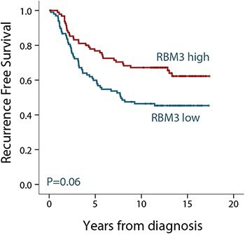
在确定RBM3可作为潜在的预后生物标志物后,研究人员进一步研究了较大乳腺癌队列中RBM3蛋白的表达1。在一组500名患有II期浸润性乳腺癌的绝经前女性患者中发现RBM3的表达与小低级别的雌激素受体(ER)阳性肿瘤相关。在分析ER阳性患者的亚组时,RBM3可作为无复发生存(RFS)的独立预测因子。如图2所示,与表达低水平RBM3的肿瘤患者相比,表达高水平RBM3蛋白的肿瘤患者的生存率有所提高。

在来自各种形式癌症的许多不同患者群组中对RBM3蛋白表达进行了进一步的分析。发现RBM3表达水平与乳腺1、结肠2、卵巢3,4、睾丸5、尿路上皮和6前列腺癌以及恶性黑素瘤7患者的生存率存在显着关联。

总结来说,RBM3在乳腺癌以及其他多种癌症中可作为优秀的预后标志物。
kaplan-meier-生存-分析

图 2.根据ER阳性乳腺癌患者中RBM3的表达而进行的无复发生存(RFS)Kaplan-Meier(生存期)分析。根据高和低的RBM3表达将患者分成两组。

RBM3抗体

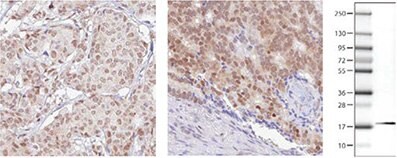
Atlas Antibodies的产品目录中有两种抗RBM3抗体:多克隆抗体HPA003624和PrecisA Monoclonal单克隆抗体AMAb90655。单克隆抗RBM3抗体AMAb90655在人类细胞系的Western Bot分析中显示出优异的特异性,并可常规用于在IHC中对福尔马林固定的石蜡包埋组织进行染色(图3)。
RBM3抗体

图 3.使用AMAb90655 对乳腺癌(左)和前列腺癌(右)中RBM3表达的免疫组化分析显示肿瘤细胞中的核阳性。WB图像显示使用AMAb90655在人内细胞系RT4裂解物中与预期相符的17 kDa条带。

参考文献

1.
Jögi A, Brennan DJ, Rydén L, Magnusson K, Fernö M, Stål O, Borgquist S, Uhlen M, Landberg G, Påhlman S, et al. 2009. Nuclear expression of the RNA-binding protein RBM3 is associated with an improved clinical outcome in breast cancer. Mod Pathol. 22(12):1564-1574. https://doi.org/10.1038/modpathol.2009.124
2.
Hjelm B, Brennan DJ, Zendehrokh N, Eberhard J, Nodin B, Gaber A, Pontén F, Johannesson H, Smaragdi K, Frantz C, et al. 2011. High nuclear RBM3 expression is associated with an improved prognosis in colorectal cancer. Prot. Clin. Appl.. 5(11-12):624-635. https://doi.org/10.1002/prca.201100020
3.
Ehlén Å, Brennan DJ, Nodin B, O'Connor DP, Eberhard J, Alvarado-Kristensson M, Jeffrey IB, Manjer J, Brändstedt J, Uhlén M, et al. 2010. Expression of the RNA-binding protein RBM3 is associated with a favourable prognosis and cisplatin sensitivity in epithelial ovarian cancer. Journal of Translational Medicine. 8(1):78. https://doi.org/10.1186/1479-5876-8-78
4.
Ehlén Õ, Nodin B, Rexhepaj E, Brändstedt J, Uhlén M, Alvarado-Kristensson M, Pontén F, Brennan DJ, Jirström K. 2011. RBM3-Regulated Genes Promote DNA Integrity and Affect Clinical Outcome in Epithelial Ovarian Cancer. Translational Oncology. 4(4):212-IN1. https://doi.org/10.1593/tlo.11106
5.
Boman K, Segersten U, Ahlgren G, Eberhard J, Uhlén M, Jirström K, Malmström P. 2013. Decreased expression of RNA-binding motif protein 3 correlates with tumour progression and poor prognosis in urothelial bladder cancer. BMC Urol. 13(1): https://doi.org/10.1186/1471-2490-13-17
6.
Jonsson L, Gaber A, Ulmert D, Uhlén M, Bjartell A, Jirström K. 2011. High RBM3 expression in prostate cancer independently predicts a reduced risk of biochemical recurrence and disease progression. Diagn Pathol. 6(1): https://doi.org/10.1186/1746-1596-6-91
7.
Jonsson L, Bergman J, Nodin B, Manjer J, Pontén F, Uhlén M, Jirström K. 2011. Low RBM3 protein expression correlates with tumour progression and poor prognosis in malignant melanoma: An analysis of 215 cases from the Malmö Diet and Cancer Study. Journal of Translational Medicine. 9(1):114. https://doi.org/10.1186/1479-5876-9-114

颗粒蛋白

颗粒蛋白是一种从单一前体蛋白质切割下来的分泌糖基化肽家族。信号肽的切割可产生成熟的颗粒蛋白,其可以进一步切割成多种活性肽。这些裂解产物被命名为颗粒蛋白A、颗粒蛋白B、颗粒蛋白C等。肽和完整的颗粒蛋白均可调节细胞的生长。颗粒蛋白家族的不同成员可作为抑制剂、刺激剂或对细胞生长具有双重作用。颗粒蛋白家族成员在正常发育、伤口愈合和肿瘤发生中是重要的[由RefSeq提供, Jul 2008]。

在Elkabets等人的论文中,通过研究将GRN表达的骨髓细胞招募到小鼠中的反应性肿瘤中来研究GRN表达在反应性肿瘤发作中的作用1。某些肿瘤可以促进位于远端解剖部位的其他肿瘤或转移细胞的生长,这被称为肿瘤诱发。在这项研究中,经严格培养的人类乳腺癌细胞被植入到小鼠体内,并显示这些细胞刺激了其他致瘤性较低的惰性转化细胞的生长。GRN被鉴定为诱发骨髓细胞中最为上调的基因。表达GRN的细胞诱导驻留的成纤维细胞表达促进恶性肿瘤进展的基因。也存在关于抗癌疗法是否可能涉及靶向GRN或活化的GRN表达细胞,从而破坏促进癌症进展的这些通信细胞系的推测。

通过使用抗GRN抗体HPA028747分析来自一组乳腺癌患者的肿瘤组织,显示高GRN表达与最具攻击性的三阴性、基底样肿瘤亚型相关并可降低患者生存率(图4)。

参考文献

1.
Elkabets M, Gifford AM, Scheel C, Nilsson B, Reinhardt F, Bray M, Carpenter AE, Jirström K, Magnusson K, Ebert BL, et al. 2011. Human tumors instigate granulin-expressing hematopoietic cells that promote malignancy by activating stromal fibroblasts in mice. J. Clin. Invest.. 121(2):784-799. https://doi.org/10.1172/jci43757
grn-表达

图 4.使用抗体 HPA028747证明GRN的表达与侵袭性肿瘤亚型相关并可降低乳腺癌患者的存活率。左图显示了每种类别(三阴性[TN] /基底或非基底)具有高GRN阳性率,而右侧的Kaplan-Meier分析则显示GRN阳性(绿色)或GRN阴性(蓝色)表达和生存期之间的相关性。
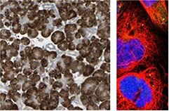
Granulin Antibodies

在Atlas Antibodies的产品目录中,有两种多克隆抗GRN抗体:HPA008763HPA028747

人-胰腺-组织-的-ihc-染色

使用抗GRN抗体(HPA008763)对人胰腺组织进行IHC染色,在外分泌腺细胞中显示出强烈的胞浆阳性。ICC-IF显示A-431细胞中囊泡的阳性。
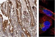
使用-抗-grn-抗体-的-ihc-分析

使用抗GRN抗体HPA028747的IHC分析显示在正常十二指肠组织中腺体细胞的强胞浆阳性和U-251MG细胞中的囊泡阳性。

Anillin

Anillin作为微丝的亚基是一种肌动蛋白结合蛋白,而微丝是细胞骨架成分之一。Anillin在大多数细胞中表达并参与基本的细胞功能,例如运动、分裂和信号转导。对anillin表达的研究表明它在多种人类肿瘤中过表达。

Breast Cancer Anillin在乳腺癌中作为治疗预测性预后生物标志物

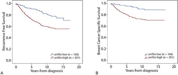
使用抗ANLN抗体HPA005680对由467例来自被诊断患有乳腺癌患者样品组成的患者队列进行Anillin表达分析。与表达低水平anillin的肿瘤患者相比,表达高水平anillin的肿瘤患者的无复发生存率(RFS)降低(图5A)。在分析乳腺癌特异性生存期时,可以看到anillin的表达与降低的生存率之间具有相同关联性(BCSS,图5B)。在O'Leary等人的一项研究中,通过Cox回归分析证实了anillin对预后的影响。在单变量和多变量分析中,高anillin表达与BCSS和RFS降低相关,并会根据肿瘤大小和等级、诊断年龄、淋巴结、ER-、PR-、HER2-和Ki67状态发生调整。

总结来说,anillin在乳腺癌中是一种较差的预后标志物
kaplan-meier-生存-分析-2

图 5.根据乳腺癌患者的aniliin表达,无复发(A)和乳腺癌特异性存活(B)的Kaplan-Meier(生存)分析。根据高和低的aillin表达将患者分成两组。

参考文献
1.
O´Leary PC, Penny SA, Dolan RT, Kelly CM, Madden SF, Rexhepaj E, Brennan DJ, McCann AH, Pontén F, Uhlén M, et al. 2013. Systematic antibody generation and validation via tissue microarray technology leading to identification of a novel protein prognostic panel in breast cancer. BMC Cancer. 13(1): https://doi.org/10.1186/1471-2407-13-175

Anillin抗体

Atlas Antibodies产品目录中有三种抗ANLN抗体:单克隆抗体AMAb90660AMAb90662,多克隆抗体HPA005680

anillin-抗体
抗-anln-抗体

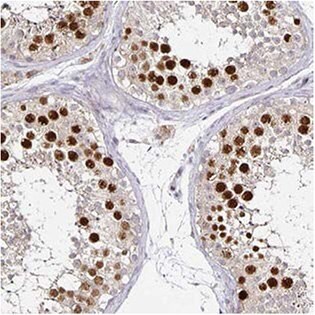
抗ANLN抗体(HPA005680)通过IHC在人睾丸生精小管细胞中显示出强的细胞核阳性。在ICC-IF中,A-431细胞的细胞核(但不是核仁)染色阳性,而在WB中抗体在RT-4和U-251 MG的细胞裂解物中检测到符合预测大小的条带。
抗-anln-抗体-2

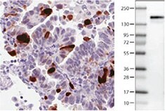
ANLN抗体AMAb90660在肺腺癌的一部分肿瘤细胞中显示出强的核免疫反应性,并且在人类细胞系U-251 MG中具有符合预测大小的条带。
抗-anln-抗体-3

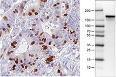
AMAb90662 ANLN抗体在结直肠癌的一部分肿瘤细胞中显示出强的核免疫反应性,并且在人U-251 MG细胞中显示出符合预测大小的条带。

登录以继续。

如要继续阅读,请登录或创建帐户。

暂无帐户?